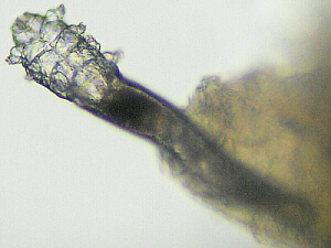
Eyelash Mite
Labeled Eyelash Mite Find these body parts on your mite and label them in your illustration.
In this lab exercise, mites in the genus Demodex will be used as
an example of organisms in class Arachnida.
Note that you will need
QuickTime
to view the movies.
The crayfish Web page provides an introduction to Phylum Arthropoda, in general, and gives characteristics of the main subphyla and classes within that phylum. That page/lab also focused more specifically on Subphylum Crustacea. In this lab, we will be focusing on Class Arachnida within Subphylum Chelicerata, and in the next lab, we will focus on Class Hexapoda within Subphylum Atelocerata.
Arachnid orders include the following. There are others; this is only a partial list of the most common ones around here. To confuse matters further, some authorities consider these groups to be subclasses with other orders within them, while other taxonomists consider these to be orders. For now, well say they are orders.
Thus, eyelash mites are classified as:
To give you a better idea of whos related to whom, if Acari is considered to be a subclass, then Order Trombidiformes (trombid = a little, timid one) is inserted under that (if Acari is considered to be an order, then Trombidiformes would be a suborder). Interestingly, Trombidiformes is the same order/suborder in which chiggers are placed, although they are in a different family within that order/suborder.
Interspecific interactions (inter = between, among) are interactions between/among organisms of different species. Any/every time organisms of different species are interacting with each other, it is considered to be some type of an interspecific interaction. Typically, these interactions are classified based on whether they are beneficial to one or both of the species involved or whether they are detrimental to one of the species involved.
Symbiosis is used to describe any relationship that involves two (or more) species living together and interacting. This is a general term which includes predation, parasitism, commensalism, mutualism, etc., but often is used, somewhat incorrectly, to mean mutualism.
Commensalism is the term used for a relationship between two species that is beneficial to one but of neutral benefit to the other. Cattle egrets follow cattle to feed on the insects stirred up by the grazing cattle. This benefits the egrets (they get food), but neither benefits nor harms the cattle.
Mutualism is used to describe a relationship between two species where both benefit. The yucca moth both pollinates and feeds on the yucca plant; acacia ants live in the thorns of, defend, and are fed by the acacia tree in which they live; and trees, in general, cant get along without mycorrhizae living in/on their roots and absorbing food for them. Many plants and their pollinators have evolved mutualistic relationships.
The relationship between eyelash mites and humans is typically one that would be called commensalism. The mites benefit from a place to live and lots of free food, while normally, the benefit to humans is of neutral value.
As mentioned above, eyelash mites are in genus Demodex (demo = people, fat; dex = an insect, a worm). There are two species that live on humans.
These microscopic mites (Demodex folliculorum and D. brevis) live in the sebaceous glands in the hair follicles not only around the eyelashes, but also on peoples noses, cheeks, foreheads, etc. One source claimed that both adults and immatures pierce epithelial cells to eat the cytoplasm, but other sources say that they feed on dead skin cells, body oils, and other leftovers that accumulate in the hair follicles. Whereas the slightly-larger females tend to stay in their hair follicle (head in, posterior end poking out), males come out, usually at night, to move around on the surface of the skin to look for mates.
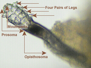
D. folliculorum measures 0.3 to 0.4 mm in length, whereas D. brevis is about half that size (0.15 [= 150 μ m] to 0.2 mm) with a similar head and thorax but a shorter abdomen. Thats amazing because these are complex, multicellular arthropods, yet are only slightly larger than a single-celled Paramecium! Like other Arachnids, they have eight legs, and it has been estimated that they can move at a rate of 8 to 16 mm/hr. Their bodies are long and somewhat scaly to help anchor them in the hair follicles. Their mouthparts (tiny chelicerae and pedipalps) resemble those of spiders, scorpions, and other arachnids, but are much smaller and designed for eating skin cells and oils within the hair follicles. Mites, like all their other arthropod relatives, have a tube-within-a-tube body plan. These mites live in the follicles, oriented parallel to the hair shaft, head inward, often with the tip of the abdomen (the opisthosoma opisth = behind, the hind part) protruding. A single follicle may contain as many as 25 D. folliculorum mites.
It has been estimated that their life cycle, from egg to adult, takes about 14.5 days, including about 5 days as an adult, and it has been reported that females may live an additional 5 days after oviposition. Interestingly, sexual maturity is reached in the larval form (called neoteny). Females remain within their hair follicles while (at night) the males wander over the skins surface from follicle to follicle in search of females. Copulation occurs at the opening of the hair follicle, after which the female crawls back into the follicle near the opening of the pilosebaceous gland (oil gland associated with a hair follicle pilos = hairy; sebum = grease, tallow) to lay her eggs.
The mites are transferred between people through facial contact (a mother kissing her baby), and it has been estimated that 96 to 98% of all people harbor these mites. In most cases, people dont even know they have these mites, but rarely (perhaps due to a suppressed immune system) the mite population can increase enough to cause skin problems, resulting in a condition known as demodicosis. A different species of Demodex causes mange in dogs.
Bibliography
anon. Demodex mite: Information from Answers.com.
http://www.answers.com/topic/demodex-mite (22-IX-2005)
anon. How do I do the test (Information for the Dermatologist or yourself). Demodex Solutions reveals the facts about demodex mites.
http://www.demodexsolutions.com/default.asp?faq.asp~mainFrame (22-IX-2005)
anon. 2001. Mystery Organism Quiz Answer - August00. BioMEDIA ASSOCIATES.
http://ebiomedia.com/feat/ansOct00.html (22-IX-2005)
Bell, Russell. 2000. The Diversity of Life.
http://www.alumni.caltech.edu/~rbell/DiversityOfLife.html (22-IX-2005)
Roque, Manolette R. and C Stephen Foster. 2005. Demodicosis. eMedicine.com, Inc.
http://www.emedicine.com/oph/topic517.htm
http://www.emedicine.com/oph/byname/demodicosis.htm (22-IX-2005)
In this lab you will be making a wet mount of material you collect from your face, but you will be using salad oil rather than water as the medium in which you will be placing your specimen. Due to the fact that these mites inhabit the oily environment in peoples pores, the use of salad oil on your slide will be a closer approximation of their natural habitat.
Some sources say that, It is quite easy to look for your own Demodex mites, by carefully removing an eyelash or eyebrow hair and placing it under a microscope, but in years of looking for them via that method, I have never had any success at finding them that way. However, you are certainly welcome to try that method. Using the method explained below, I have never had any trouble finding mites, often several on one slide.
Note: one Web page quotes a procedure, similar to the following, as explained on page 177 of
Wilson, Edward O. 1992. The Diversity of Life. W. W. Norton. New York.
Several other Web pages quote the same procedure, but neglect to reference E. O. Wilsons book as the source of that quote, while several other Web page authors use other slight variations on this method.
Obtaining samples from different face parts (forehead, nose, cheeks) may boost your chances of finding mites.
Eyelash Mite
Labeled Eyelash Mite
Find these body parts on your mite and label them in your
illustration.

Cephalothorax at Bottom Left, Legs to Right (© DBF)

Abdomen at Top (Note Segments) (© DBF)

Close-up of Abdomen

Close-up of Abdomen

Close-up of Cephalothorax, Mouthparts to Right

Close-up of Cephalothorax, Mouthparts to Right

Close-up of Cephalothorax, Mouthparts to Right

Close-up of Cephalothorax, Mouthparts to Right
Make sure you have all of the following in your lab notebook: