Lubber Grasshopper (Insecta)
Background Information
The purpose of this lab exercise is to become familiar with
the external and internal anatomy/morphology of
the Eastern Lubber Grasshopper, Romalea guttata (also known as R.
microptera.
The Eastern Lubber Grasshopper is classified as:
Kingdom Animalia
Phylum Arthropoda
Subphylum Atelocerata
Class Hexapoda
Subclass Insecta
Order Orthoptera (ortho = straight; ptera = wing)
Suborder Caelifera (= grasshoppers, not katydids or crickets)
Family Acrididae
Subfamily Romaleinae (some authors call Romaleidae a family)
Genus Romalea
Species guttata (or microptera)
 Insects exhibit one of two types of metamorphosis.
Those with gradual metamorphosis change from egg to nymph to adult.
In these insects (grasshoppers, roaches, true bugs), the nymphs look like
miniature adults without wings, usually living in the same environment and
eating the same food. Insects with complete metamorphosis go from
egg to larva to pupa to adult (larva = ghost, specter; pupa =
doll). Larvae of these insects look very different from the adults, usually
live in a totally different environment and eat different food. The pupa is
a resting stage where much transformation takes place. Probably the example
of complete metamorphosis with which most people are familiar is that of a
caterpillar (larva) changing to a chrysalis (pupa) then to a butterfly
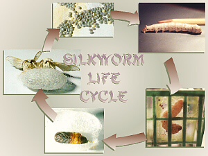
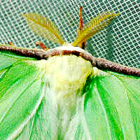
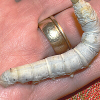
(adult). Silkworms are the caterpillars of Bombyx mori, a species
of moth. As many, but not all, moths do, they spin cocoons prior to molting
to a pupa/chrysalis, and humans have discovered that it is possible to
unwind the silk thread that makes up their cocoons and weave it into cloth.
Insects exhibit one of two types of metamorphosis.
Those with gradual metamorphosis change from egg to nymph to adult.
In these insects (grasshoppers, roaches, true bugs), the nymphs look like
miniature adults without wings, usually living in the same environment and
eating the same food. Insects with complete metamorphosis go from
egg to larva to pupa to adult (larva = ghost, specter; pupa =
doll). Larvae of these insects look very different from the adults, usually
live in a totally different environment and eat different food. The pupa is
a resting stage where much transformation takes place. Probably the example
of complete metamorphosis with which most people are familiar is that of a
caterpillar (larva) changing to a chrysalis (pupa) then to a butterfly
(adult). Silkworms are the caterpillars of Bombyx mori, a species
of moth. As many, but not all, moths do, they spin cocoons prior to molting
to a pupa/chrysalis, and humans have discovered that it is possible to
unwind the silk thread that makes up their cocoons and weave it into cloth.
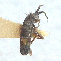
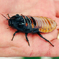
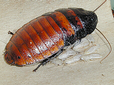 Members of Order Orthoptera have gradual metamorphosis.
Their life cycles consist of eggs, nymphs (which resemble the adults, as
well as sharing the same habitat, food, etc.), and adults. The same is true
for roaches, such as the mother and her new babies pictured here.
Members of Order Orthoptera have gradual metamorphosis.
Their life cycles consist of eggs, nymphs (which resemble the adults, as
well as sharing the same habitat, food, etc.), and adults. The same is true
for roaches, such as the mother and her new babies pictured here.
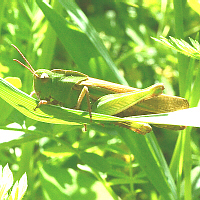
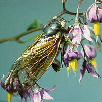
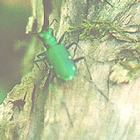
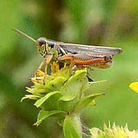
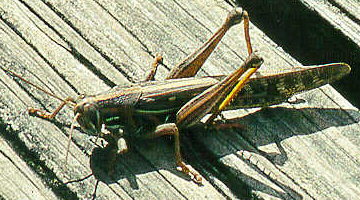 Other members of family Acrididae include other types of grasshoppers,
including the American Grasshopper pictured here.
While this American Grasshopper has long wings and can fly, the lubber
grasshoppers have short, stubby wings (hence the species name,
microptera, micro = small) and cannot fly.
Other members of family Acrididae include other types of grasshoppers,
including the American Grasshopper pictured here.
While this American Grasshopper has long wings and can fly, the lubber
grasshoppers have short, stubby wings (hence the species name,
microptera, micro = small) and cannot fly.
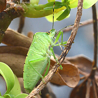
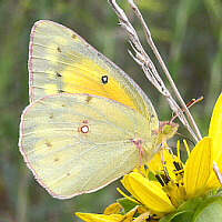
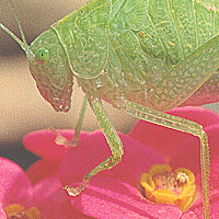
As its common name implies, the Eastern Lubber Grasshopper
occurs in the
eastern United States, more specifically the southeastern US.
Nymphs (young) are typically black with a red or yellow
stripe down the
center of their backs. Adults have one of a few typical color patterns
(phases). Some are a dull yellowish (greenish-yellow) with black markings,
others are more orangish with black markings, and yet others are black with
red or yellow stripes. Any/all of these yellow-and-black or red-and-black
color patterns serve as warning coloration (aposematism
apo
= from, off, away; sema, semato = mark, sign, signal, seal), a type
of defense mechanism that advertizes their bad taste to would-be predators.
They can also startle predators by forcing air out of their abdominal
spiracles to produce a hissing sound and by secreting a bad-smelling and
bad-tasting liquid.
Characteristics of the Orders of Class Hexapoda
Order
example(s) |
Type of Front Wing |
Type of Back Wing |
Other Notes |
Photo |
| Subclass Entognatha |
| (endo,
ento = within, inner; gnatho = the jaw), mouthparts within the head, primarily wingless, simple metamorphosis, no longer considered to be insects |
Protura
proturan |
none |
none |
(proto = first, original; ura = the tail) |
|
Collembola
springtail |
none |
none |
(coll = glue; embola
= a bolt or wedge) collophore on bottom of 1st
abdominal segm., for water uptake + furcula = jumping organ
on ventral abdomen
|
 |
Diplura
dipluran |
none |
none |
(diplo = double, two;
ura = tail) group name refers to presence of two
filaments arising from the posterior end of the body |
|
| Subclass Insecta |
| (ecto = outside,
out, outer) ectognathous mouthparts stick out from head |
| Apterygota |
| (a- = not,
without; ptero = wing, feather) primarily wingless, simple
metamorphosis |
Microcoryphia
jumping
bristletails |
none |
none |
(micro = small;
corypha = head, top) body more cylindrical than silverfish, small
head with large compound eyes, body covered with scales |
|
Thysanura
silverfish
firebrats |
none |
none |
(thysan = fringe) somewhat
flattened body, three taillike structures on posterior end, body often
covered with scales
|
|
| Pterygota |
| winged (a few are secondarily wingless) |
| Exopterygota |
(exo
= out, outside) gradual metamorphosis, wing pads develop externally,
young are called nymphs (naiads if aquatic)
Ephemeroptera, Odonata, and Plecoptera, which have aquatic
naiads,
are said to be hemimetabolous
(hemi = half).
|
Ephemeroptera
mayflies |
membranous |
membranous, smaller than front wings |
(ephemer = for a day,
temporary) aquatic naiads, winged subimago, then adult; very
short-lived as adults |
|
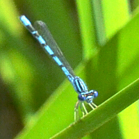
Odonata
dragonfly
damselfly |
membranous
long & narrow |
membranous
long & narrow |
(odonto = tooth) have teeth
on mandibles, aquatic naiads; chewing mp; long & slender
|
 |
Grylloblattaria
rock crawlers |
|
|
(gryll = cricket; blatta
= cockroach) rare, nocturnal insects found in cold places like the edges
of glaciers |
|
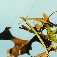
Phasmida
walkingstick
leaf insect |
leathery tegmina
(or absent) |
membranous
(or absent) |
(phasmato = apparition,
phantom) chewing mp; look like sticks or leaves, &
well-camouflaged |
 |
Orthoptera
grasshopper
katydid
camel cricket
cricket |
leathery tegmina
(or absent) |
membranous
(or absent) |
(ortho = straight)
jumping back legs; chewing mouthparts |
 |
Mantodea
mantis |
leathery tegmina
(or absent) |
membranous
(or absent) |
(manti, mantid,
mantis = a soothsayer, a kind of grasshopper) chewing mp; front
legs adapted for catching prey |
 |
Blattaria
cockroach |
leathery tegmina
(or absent) |
membranous
(or absent) |
(blatta = cockroach)
chewing mp; legs adapted for running |
 |
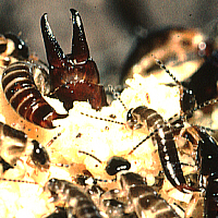
Isoptera
termite |
membranous
(or absent) |
membranous; same size as front (or absent) |
(iso = equal) light-colored;
no waist; chewing mp; small size;
social with castes, winged reproductives; |
|
Dermaptera
earwig |
shortened = brachypterous
(or absent);
leathery, called tegmina or elytra |
membranous; folded under front wings (or absent) |
(derm = skin) forceps-like
cerci at end of abdomen |
 |
Embiidina
webspinners |
|
|
(embi = lively, long-lived)
small; tropical & subtropical; silk glands in basal segment of
front tarsus |
|
Plecoptera
stoneflies |
membranous |
membranous, bottom area folded under at rest |
(pleco = twine, twist,
braid, twisted, folded) aquatic naiads |
|
Zoraptera
zorapterans |
membranous or none |
membranous or none |
(zoro = alive, living, pure,
strong; a- = not, without) only wingless ones known
when order was named; tiny; gregarious;
|
|
Psocoptera
psocids |
membranous or none |
membranous or none |
(psoco = rub small) indoor
in books = booklice; outdoors = barklice; small & soft-bodied;
order name from gnawing habits |
|
Phthiraptera
lice
Suborder Mallophaga
chewing lice;
Suborder Anoplura
sucking lice |
none |
none |
(phthir = lice)
ectoparasites (mostly on birds or mammals) |
|
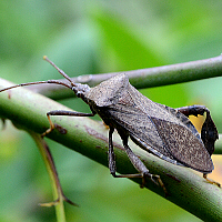
Hemiptera
true bug |
half-leathery, half-membranous hemelytra;
X when folded |
membranous |
(hemi = half)
piercing-sucking mp; |
 |
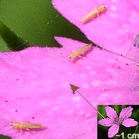
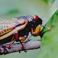
Homoptera
leafhopper
cicada
aphid
scale insect |
membranous
(or absent) |
membranous
(or absent) |
(homo = same,
like, alike) piercing-sucking mp |
 |
| similar
to each other (unlike true bugs), held
rooflike or tentlike over body when at rest |
| |
Thysanoptera
thrips |
2 larval instars with internal wing development followed by 3rd or 3rd & 4th instars that are quiescent (prepupa & pupa) & have external wing pads; adults with or without wings |
(thysano = fringe)
metamorphosis intermediate between gradual and complete; pupa sometimes with cocoon; tiny size |
|
| Endopterygota |
(endo = within, inner) complete metamorphosis,
wing pads develop internally until pupal stage, young called larvae |
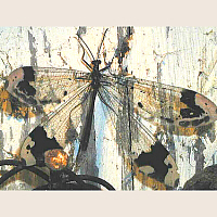
Neuroptera
dobsonfly
lacewing
antlion |
membranous |
membranous |
(neuro = nerve, sinew, cord)
named for wing veins; dobsonfly larvae are aquatic; many prey
on other insects
(Photo © D.B.
Fankhauser) |
 |
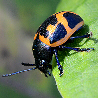
Coleoptera
beetle |
hard, shell-like elytra |
membranous |
(coleo = a sheath) chewing
mouthparts; largest order with ~40% of all insects |
 |
Strepsiptera
twisted-wing
insect |
males winged, females wingless |
males winged, females wingless |
(strepsi = twisted, a
turning or twisting) tiny; males free-living; females parasitic on
other insects, may be legless |
|
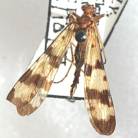
Mecoptera
scorpionfly |
membranous |
membranous |
(meco = long, length) tip
of males abdomen curls up, resembling shape of scorpions (photo is female);
long, snout-like head |
 |
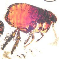
Siphonaptera
flea |
none |
none |
(siphon = tube, pipe)
pupa in cocoon; blood-sucking; jumping; small & flat |
 |
Diptera
fly
mosquito
cranefly
gnat |
membranous |
modified as halteres |
(di = two) adults with
sponging, cutting-sponging, or piercing-sucking mp |
 |
Trichoptera
caddisflies |
membranous, hairy, may be scaly |
membranous, hairy, may be scaly |
(tricho = hair) larvae
are aquatic, making protective cases of silk + stones, bits of leaves,
etc. |
|
Lepidoptera
butterfly
skipper
moth |
bright color due to scales |
bright color due to scales |
(lepido = a scale) siphoning mp in adults, chewing in larvae (caterpillar) |
 |
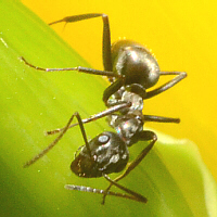
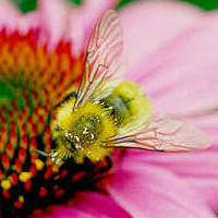
Hymenoptera
bee
ant
wasp |
membranous
(or absent) |
membranous,
smaller than front
(or absent) |
(hymeno = a membrane) have a
waist; chewing mp; many can sting; many social in colonies; often black
& yellow bodies |
 |
Lubber Grasshopper Anatomy and Dissection:
External Anatomy
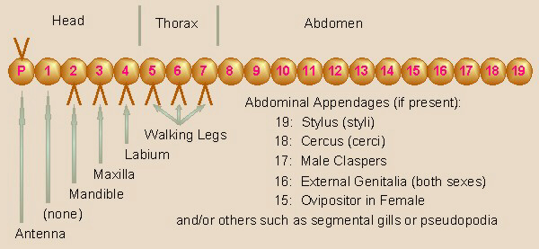
First, a review of the locations of generic insect segments
and appendages would be useful. In this diagram, the segment labeled as #8
would be the first abdominal segment (labeled below as A1) and, in
grasshoppers only (not true for other kinds of insects), bears
the tympana (sing. = tympanum), #9 would be the second
abdominal segment (labeled as A2), and #10 through 14 would correspond to
A3 through A7. The segment labeled as #15 would be A8, and in female insects,
bears the first half of the ovipositor. The segment labeled as #16
would be A9 and in females, bears the second half of the ovipositor,
while in males bears portions of the male genitalia. The segment labeled
as #18 corresponds to A11 and bears the cerci (sing. = cercus) in both sexes,
while #19 is A12 and bears the styli (sing. = stylus) in males. All of
these structures are discussed below.
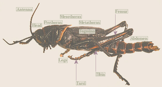
Observe the external
anatomy of your grasshopper. Note that an insects body is divided into
three main sections: head, 3-segmented thorax, and abdomen.
On your grasshopper, find all body parts identified in this illustration.
Draw your grasshopper, and label those body parts on your
drawing. Optionally, if available, view (and draw) pinned specimens of
other insects, and find these body parts on those insects, too (caution:
pinned insect specimens are very fragile and easily broken if not handled
gently).
Body Coloration: Mimicry and Camouflage
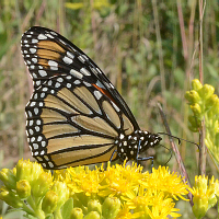
Monarch Butterfly
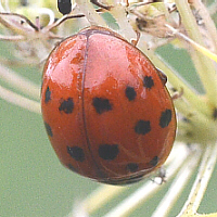
Ladybird Beetle
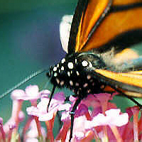
As mentioned above, a number of dangerous or distasteful insects exhibit
red-and-black or yellow-and-black stripes as warning coloration
(aposematism apo = from, off, away; sema, semato =
mark, sign, signal, seal) to warn of their bad taste, stingers, etc.
to deter would-be predators. Both Monarchs and ladybird beetles (ladybugs)
taste bad.
 |
 |
 |
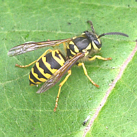
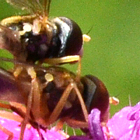
| Yellow Jacket Wasp Model |
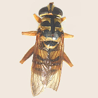
Flower Fly Mimic |
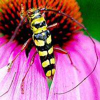
Locust Borer Beetle Mimic |
Other edible, non-venomous insects have taken advantage of
those bright color patterns, using mimicry to make potential predators
think they, also, taste bad or are dangerous. For example, its a big
mistake to tangle with a yellow jacket, but Locust Borers and flower flies
are pretty benign. Yet, due to their mimicry, theyre probably a lot less
likely to be eaten.
Another, often-cited example is Viceroy butterflies mimicking Monarchs, which
are known to taste bad, but it turns out that Viceroys, too, may not taste
all that good.
 |
 |
 |
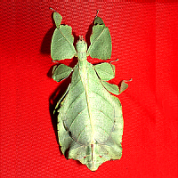
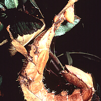
| Female Javanese Leaf Insect |
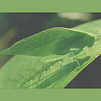
Female Greater Angle-wing Katydid |
Dead Leaf Butterfly |
Other insects depend on not being seen (camouflage) to
protect themselves from predators. Our local North American Walkingstick is
a good example, as is its relative, the Javanese Leaf Insect. Many species
of katydids and several kinds of butterflies also closely resemble leaves.
This dead leaf butterfly has its head down and to the right. What looks
like the petiole (stem) of the leaf on the upper left is actually pointed
tips on the hind wings. Look carefully and you may be able to see several
of its legs and its antennae.
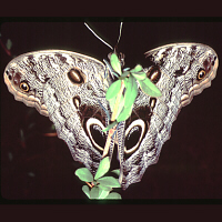
Owl Butterfly © M. K. Busching
Now, suppose youre an insectivore (an animal that eats insects), walking
through the tropical rainforest at dusk looking for some supper. Hmmm... it
smells like an insect, so lets go check it out. Eeek! Theres an owl
out there, and its getting ready to eat ME! Just look how big its eyes
are! Forget supper, Ive gotta get outta here!
Well, chalk one up for the Owl Butterfly. Owl Butterflies sit with their
wings shut, and so are fairly well camouflaged in the dappled light of the
rainforest. When disturbed/alarmed, they suddenly open their wings,
displaying the large eyespots that looks a whole lot like owls eyes. The
centers of the wings and the body add to the realism by appearing to be
the beak of the owl.
Examine your grasshopper
paying special attention to its color pattern. Pokeweed is reported to be
on lubbers list of favorite foods, and they, like many other insects,
are able to sequester/store the toxic chemicals from pokeweed and the other
plants they consume somewhere in their bodies (the exoskeleton is a common
site for such storage).
What does your grasshoppers coloration tell you about it into which of
the above-mentioned categories does it fall?
A more detailed discussion of each body region (head, thorax,
and abdomen)
and its characteristics and appendages follows.
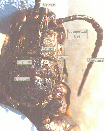
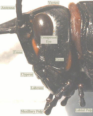
Head
The top of the head is the vertex.
The frons is the face area, bounded by the antennae and the
top of the mouthparts. The gena is the cheek region below the
eye and above the top, side edges of the mouthparts.
The ventral portion of the frons is known as the clypeus.
Below and attached to the bottom of the clypeus is a heavy flap, the
labrum, which functions as the upper lip. Behind the maxillae is
the labium, serving as the lower lip,
a complex structure formed by the fusion of the paired second maxillae.
More information on the mouthparts is included below.
Find all these parts on your grasshoppers head, then draw in your lab
notebook and label them
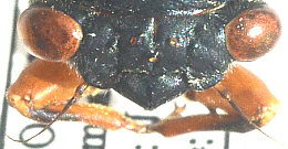
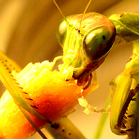
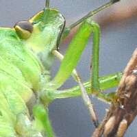
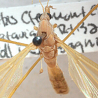
Insect Eyes
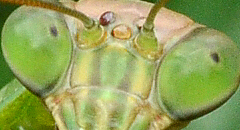
 Compound eyes The compound eyes are composed of many, small,
hexagonal facets, called ommatidia (ommato = the eye). Each of these is a separate
eye and motion is detected by the interplay of light and dark across the
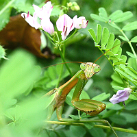
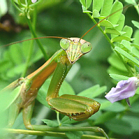
ommatidia. This photo is of the left compound eye on a female Chinese
Mantis. Her center
ocellus is also visible as a small brown bump to the left of the
forward-most antenna.
Compound eyes The compound eyes are composed of many, small,
hexagonal facets, called ommatidia (ommato = the eye). Each of these is a separate
eye and motion is detected by the interplay of light and dark across the
ommatidia. This photo is of the left compound eye on a female Chinese
Mantis. Her center
ocellus is also visible as a small brown bump to the left of the
forward-most antenna.
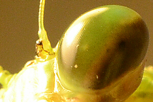
 The ommatidia are oriented such that their bottom ends come together and
often appear to
form a false pupil, visible through the compound eyes, as is seen in
this mantis eyes. Because the false pupil indicates location of the
bottom ends of the
ommatidia, down in the center, inside of the eyes, if you were really looking
at this guy in person, his false pupils would appear to change position as
you move your head. The false pupil helps the
insect to see better. Also, notice the three brown spots between his
antennae. These are individual eyes called ocelli (sing. = ocellus;
ocellus = a little eye)
which are discussed next.
The ommatidia are oriented such that their bottom ends come together and
often appear to
form a false pupil, visible through the compound eyes, as is seen in
this mantis eyes. Because the false pupil indicates location of the
bottom ends of the
ommatidia, down in the center, inside of the eyes, if you were really looking
at this guy in person, his false pupils would appear to change position as
you move your head. The false pupil helps the
insect to see better. Also, notice the three brown spots between his
antennae. These are individual eyes called ocelli (sing. = ocellus;
ocellus = a little eye)
which are discussed next.
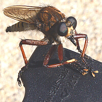
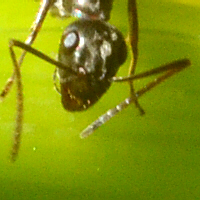
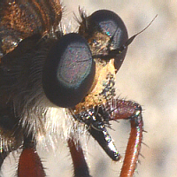
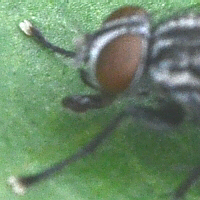
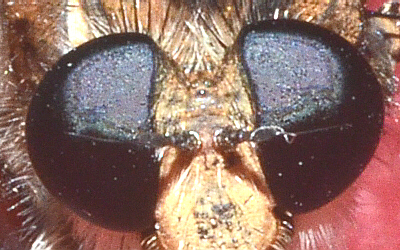 Ocelli Many, but not all, insects also have three individual
ocelli in between the compound eyes. This robber flys ocelli are
visible in the trough/valley between his/her compound eyes. Also, note the numerous facets of
the flys compound eyes.
Ocelli Many, but not all, insects also have three individual
ocelli in between the compound eyes. This robber flys ocelli are
visible in the trough/valley between his/her compound eyes. Also, note the numerous facets of
the flys compound eyes.
 This periodical cicadas three ocelli are the three, small, brown spots
between the compound eyes.
This periodical cicadas three ocelli are the three, small, brown spots
between the compound eyes.
Use a dissecting scope to
observe your grasshoppers eyes. Can you see the individual ommatidia?
Also look carefully at the front of the vertex and the region where the
vertex and the frons join. Does your grasshopper have ocelli?
Insect Antennae
 Antennae (antenna = a sailyard) are modified appendages serving a sensory
function. Besides touch receptors, most insects also have chemoreceptors
(sense of smell) in/on their antennae. If available, observe and compare
antennae on various other types of insects.
Antennae (antenna = a sailyard) are modified appendages serving a sensory
function. Besides touch receptors, most insects also have chemoreceptors
(sense of smell) in/on their antennae. If available, observe and compare
antennae on various other types of insects.
| Type |
Description |
Photo |
| aristate |
(arista = an awn, bristle)
antenna often with 3 segments and having a filamentous arista
coming out of the side of the last/tip segment
typical of many
kinds of flies
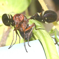
These two little flies are mating (male on top). In this photo,
their abdomens are off the picture to the left, and their heads are
facing to the right. The antennae are yellow lumps off the fronts of
their heads, and the aristae are the black filaments sticking out of
them. |
 |
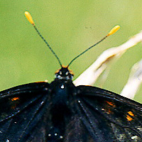
| capitate |
(capit = the head)
thinner antenna suddenly widening at the end into an enlarged, headlike
tip
typical of butterflies, skippers, and a number of other types
of insects |
 |
| clavate |
(clava = a club) antenna
gradually widening at the end into an enlarged or clublike tip
|
|
| filiform |
(fili = a thread) the
whole antenna hairlike or threadlike, often longer in overall length
typical of many types of insects |
 |
| flabellate |
(flabella = a fan) each
segment enlarged on one side into a platelike or leaflike projection,
giving the whole antenna the appearance of a fan
|
|
| geniculate |
(genicul = the elbow, knee)
the basal segment is elongated, and the other segments held at an angle
to that segment, giving the antenna an overall elbowed appearance
typical of ants, bees, and many other Hymenopterans |
 |
| lamellate |
(lamell = a small plate) a thinner antenna with a few of the tip segments enlarged into platelike or leaflike projections
typical of Scarab beetles |
 |
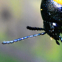
| moniliform |
(monil = a necklace, string
of beads) each segment of the antenna rounded and beadlike
This Milkweed Leaf Beetles antennae really are only half-way moniliform,
and arent really rounded into totally bead-like shapes. |
 |
| pectinate |
(pectin = a comb) each
segment with one or two (bipectinate) lateral projections giving the
antenna an overall comblike appearance
typical in some kinds of
moths and a few other kinds of insects |
 |
| plumose |
(plumo = a feather) each
segment with numerous, filamentous projections, giving an overall
feathery appearance
typical in male mosquitoes and some other
kinds of insects |
|
| serrate |
(serrat = a saw) antenna
with each segment pointed on one side, giving an overall appearance that
the antenna is toothed along the edge like a saw
|
|
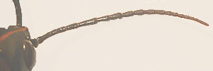
| setateous |
(seta = a bristle) the
whole antenna thin and bristlelike, shorter in length
typical in
dragonflies, damselflies, and cicadas |
 |
| stylate |
(stylo = a pillar, stake,
column, a pointed instrument) antenna often 3-segmented, with the tip
(not side) of the last segmented becoming/bearing a thin, threadlike
style
typical in some kinds of flies |
 |
Examine your grasshoppers
antennae. Based on this list and photos (above), which type of antennae
does the grasshopper have? If not already part of your drawing of the
grasshoppers head, draw your grasshoppers antennae. Include a label
telling which kind of antennae these are.
Insect Mouthparts
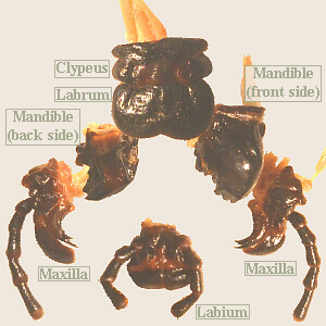 Mouthparts There are four types, four layers, of mouthparts.
These are listed here from front to back. The labrum is not
considered to be an appendage, but the mandibles, maxillae, and labium are
appendages of segments 2, 3, and 4, as shown in the diagram, above.
Mouthparts There are four types, four layers, of mouthparts.
These are listed here from front to back. The labrum is not
considered to be an appendage, but the mandibles, maxillae, and labium are
appendages of segments 2, 3, and 4, as shown in the diagram, above.
- Labrum (labr = a lip) A flap covering the front of the
mandibles. The labrum is attached to and hangs down from the
clypeus.
- Mandibles Heavy, tooth-like appendages modified for chewing.
Note that these are not homologous to our teeth note the
sideways motion. The mouth lies between and above these.
- Maxilla (sing.)/Maxillae (pl.) (maxill = jaw,
jawbone) Also modified appendages, these are found immediately behind
the mandibles. The outer, thread-like portion is the
maxillary palp (pl. = palpi) (palp = touch, feel).
- Labium (labi = a lip) This is the last of the
mouthparts. Modified appendages from each side are fused into one piece.
The outer, thread-like portions are the labial palps.
While chewing mouthparts are considered the primitive or
ancestral form, in many other types of insects, including true bugs,
butterflies, etc., the same four structures are present, but may be highly
modified for other diets and methods of feeding. Some of these types are
listed below.
Carefully remove each
mouthpart from at least one side (left or right both is OK, too) of the
head/mouth by using a forceps to pull it out from the base in order to observe
it more closely. As you remove each part, first examine it carefully to see
how it fits and to better view the remaining mouthparts. Then, after each
is removed, use a dissecting scope to get a better idea of what that
mouthpart looks like. Draw that mouthpart, labeling its sub-parts (for
example, the palpi.
Once you familiarize yourself with the mouthparts of the grasshopper, also
view and draw the mouthparts of any other insects that may be available. In
addition to chewing mouthparts, also look for insect specimens with other
types of mouthparts.
| Type |
Description |
Photo |
| mandibulate, chewing |
grasshoppers, roaches, mantises,
caterpillars, wasps
this is thought to be the most primitive form from which all the others
are derived |
 |
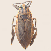
| piercing-sucking, haustellate |
mosquitoes, true bugs, cicadas,
robber flies
these can pierce the host tissue (plant or animal) and suck up fluids;
may secrete digestive enzymes, first, to liquify solid food;
in true bugs and cicadas, the mouthparts are collectively called a
beak or proboscis |
 |
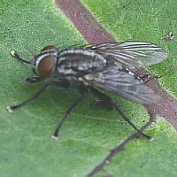
| sponging |
house fly
if needed, flies
will first secrete digestive enzymes to liquify their food, then all
food, in a liquid form, is sponged up by the mouthparts |
 |
| cutting-sponging |
horse fly
these cut a hosts skin, then sponge up the blood that comes out (this one wouldnt hold still long enough for a mouth picture) |
 |
| proboscis, siphoning |
butterflies and moths
mouthparts are called a proboscis and function like a soda
straw |
 |
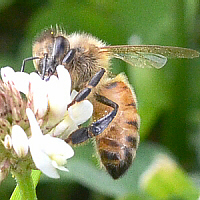
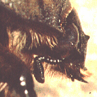
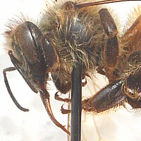
| chewing-lapping |
honey bees
bees need to chew on honeycomb to shape it, and also lap up nectar/honey;
scissor-like mandibles can be seen at the base/bottom of the head, on
the left, while the long tongue sticking out is used to lap up honey |
|
Thorax
The thorax includes 3 body segments: the prothorax,
mesothorax, and metathorax. Each of these segments bears a
pair of legs. Additionally, the meso- and metathorax frequently each
bear a pair of wings (which are not considered to be
appendages).
- Prothorax (pro = before, in front of) Bears one pair
of walking legs, no wings in any insects.
- Mesothorax (meso = middle) Bears one pair of walking
legs. In many insects, one pair of wings is also present. These are not
homologous to bird wings they are not modified appendages, but
rather merely flaps of exoskeleton. Frequently these front wings are
modified as covers for the hind wings, and in the case of grasshoppers,
these narrow, leathery wing covers are called tegmina. Lubbers
wings are short (brachypterous brachy = short; ptero
= wing, feather), and they cannot fly.
Refer to the chart, below, which describes the main types of wings.
- Metathorax (meta = between, with, after) Bears one
pair of walking legs, and in many insects, one pair of wings (modified as
halteres (halter = a leaping weight) in Order Diptera
flies and their relatives).
Each thoracic segment has a sclerotized (hardened;
sclero = hard) dorsal portion, the notum, (nota,
notum = the back) specifically pronotum, mesonotum, and metanotum
and a sclerotized ventral portion, the sternum, (stern =
breast, breastbone) joined by a lateral membranous area.
Compare the flexibility of the thorax versus the abdomen.
Why do you suppose this is? (Hint: To where do you think the muscles used
in walking attach?)
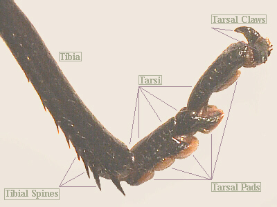
Insect Legs
 The parts of an insect leg are named such that the locations of these
parts roughly correspond to the locations of the parts of the human leg.
The small segment that attaches to the body is the coxa (coxa
= hip), followed by another small segment, the trochanter
(trocho = wheel, disk in reference to the ball at the top of the
femur in humans), then the larger femur (femur = thigh), the
tibia (tibia = flute, the shin bone) and finally, the series
of smaller segments farthest from the body are the tarsi
(tarsus = the ankle).
The parts of an insect leg are named such that the locations of these
parts roughly correspond to the locations of the parts of the human leg.
The small segment that attaches to the body is the coxa (coxa
= hip), followed by another small segment, the trochanter
(trocho = wheel, disk in reference to the ball at the top of the
femur in humans), then the larger femur (femur = thigh), the
tibia (tibia = flute, the shin bone) and finally, the series
of smaller segments farthest from the body are the tarsi
(tarsus = the ankle).
On the ventral surfaces of the tarsi, note the whitish (or at least, lighter
colored) tarsal pads. These suction cups are controlled by hydraulic
pressure within the animals body and serve to cling to objects. Also note
the tarsal claws on the last segment. Different insect species have
from two to five tarsal segments per leg and some have differing numbers of
tarsi on the pro-, meso-, and metathoracic legs.
| Type |
Description |
Photo |
| cursorial: running |
cockroaches, tiger beetles
(curso = run, a runner) |
 |
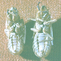
| fossorial: digging |
mole cricket, cicada nymphs
(-fossor = a digger)
As can barely be seen on these cicada skins, the front legs of cicada
nymphs are modified for digging. |
 |
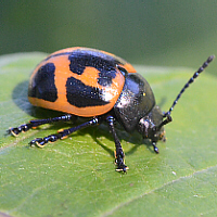
| gressorial: walking (vs cursorial = running) |
grasshopper front legs, milkweed leaf beetle
(gress = walk, walking) |
 |
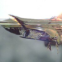
| natatorial: swimming |
diving water beetle, giant water
bug, waterboatman
(natan = swimming)
Notice how especially the hind leg on this giant water bug is broad and
oar-like for swimming. The front legs, by the way, are raptorial. |
 |
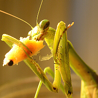
| raptorial: grasping prey |
mantis, water strider
(raptor = seize, plunder, a plunderer)
Notice how Mom Mantis can fold her tarsi back out of the way to protect
them while manipulating her prey/food. |
 |
| saltatorial: jumping |
grasshopper, katydid, camel cricket,
cricket
(salta = leap, dance)
This delicate little lady is a tree cricket. |
 |
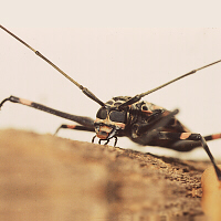
| scansorial: climbing |
Harlequin Beetle
(scansor = climb)
elongated joints with large tarsal claws; female Harlequins legs are a
bit
shorter than those of the male; when mating, the male must have front
legs that are long enough to reach around the female and grab hold of
the tree trunk. |
 |
| Other Types |
pollen basket
pollen rake
etc. |
Honey bees legs are highly specialized
for a number of important functions related to gathering pollen. See below
for a labeled and annotated illustration. |
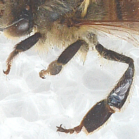 |
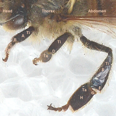
- Ti = tibia
- Ta = first tarsal segment
- A = eye brush (a honey bee moistens its forelegs with its
tongue, then brushes all the pollen from its head and body back to
the hind legs)
- B = notch in the top of the first tarsal segment is used in
cleaning the antennae
- C = pollen brush
- D = wax spine (the undersides of the bees abdominal segments
secrete wax, and it is thought that this spine at the end of the
mesothoracic tibia helps to remove the wax from the abdomen as it
forms)
- E = pollen basket or corbicula (corbi = a basket;
-cula = little on the outside of the
hind tibia, a shiny cavity, into which pollen is placed, surrounded by
a fringe of hairs; a hair poking through the middle of the pollen
load secures it, and honey/nectar is used to glue it together)
- F = pollen rake (this fringe of spines on the tibia, along
with the auricle on the first tarsal segment work together to compress
the pollen
thats transferred there from the pollen comb, and then the compressed
pollen is transferred to the pollen basket to be carried)
- G = auricle (works together with rake to pack pollen
together)
- H = pollen comb (on inner surface not visible, here; pollen
is transferred to pollen comb, then compressed and transferred to the
pollen basket)
|
| pulvillus (pulvilli), a type of
tarsal pad |
Many flies (Order Diptera) have a
pair of pads attached at the bases of the tarsal claws. In this fly,
they are visible as the pair of white pads on the end of each foot.
|
 |
| fruit fly male sex comb |
Male fruit flies have a black
sex comb on their first prothoracic tarsal segment. These are used
to grab the females abdomen and/or wings as part of their courtship prior
to copulation. |
|
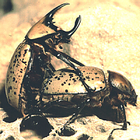
| suction disc on predaceous
diving beetles |
Not visible in this picture, but
male diving beetles (Dytiscidae) have large suction discs on their
front tarsi. These are used to hold onto the smooth, shiny elytra of the
female during mating. Also notice that the hind legs (one sticking up in
the photo) are flattened and
fringed with hairs to serve as oars, enabling the beetles to swim.
Note the air bubble (most of which is under the elytra and not visible),
portions of which are visible, giving the body a silvery color.
|
 |
tympanum on katydids
and crickets |
Katydids and crickets that sing
have a tympanum (tympanum = a drum) on the tibiae of their
front legs. This organ enables them to hear. In this photo, this
katydid nymphs tympanum is the brown spot just below her front knee.
|
 |
Examine and draw your
grasshoppers legs. How are the grasshoppers three pairs of legs similar,
and how are they different? How many tarsi does a lubber have on each of
its legs (make sure your drawing is accurate)?
As time and specimens
allow, observe legs on a variety of
pinned insects. Look for and draw legs from as many of the following
categories as are available to examine.
If available in lab or
later, while your class is on a field hike, observe a live insect walking.
Note that it
moves its legs in sets of three (front and back on one side and middle of
opposite side) so that it is always resting on a tripod (the most sturdy
and secure base of support).
Insect Wings
Insect wings are modified in a number of ways, as listed in
this table.
| Type |
Description |
Photo |
| apterous |
(a- = not, without)
This refers to any insect in which wings are lacking. Again, this occurs
in insects in many groups where close relatives have full-length wings,
and in that case, such insects would be called secondarily apterous.
In other insect groups, such as whole orders whose members have never
had wings, they are said to be primarily apterous.
|
 |
| brachypterous |
(brachy = short)
This refers to an insect that has short wings it occurs in insects in
many groups where close relatives have full-length wings. Since
brachypterous
refers to the length of the wings, it may be combined with other terms
such as elytra or tegmina.
(photograph of female Australian
Walkingstick © M. K. Busching, retired Curator of Invertebrates at the
Cincinnati Zoo)
|
 |
| elytra |
beetles
(elytr = a sheath, cover)
the hardened forewings characteristic of Order Coleoptera |
 |
| halteres |
flies, mosquitoes, crane flies
(halter = a leaping weight)
hind wings modified as knob-like balancing structures characteristic of
Order Diptera, and since they are often tiny and not easily visible,
dipterans appear to have only two wings |
 |
| hemelytra |
true bugs
(hemi = half)
wings whose close (proximal) portion is leathery and whose farther
(distal) portion is membranous, and which are held such that they form
an X over the insects back characteristic of Order Hemiptera |
 |
| membranous |
honey bees, dragonflies
membranous wings are typical of a wide variety of insects |
 |
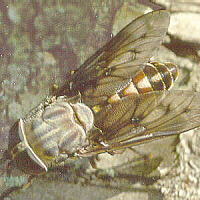
| pictured wings |
typical especially in some types of
flies (Diptera) which have spots or bands on their wings also true of
some types of dragonflies
Picture-wing flies often use their wings in a signaling display as part
of their courtship ritual.
|
 |
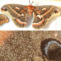
| scale-covered wings |
butterflies, moths
wings that, underneath, are membranous, but they are covered with scales,
which may be brightly colored characteristic of order Leipdoptera;
some members of Order Trichoptera (caddisflies) also have scales on their
wings
This photo shows a Cecropia Moth, a very large, local moth, and a close-up
of a portion of one of her wings, showing the scales. |
 |
| tegmina |
grasshoppers, katydids, crickets,
cockroaches, walkingsticks, mantises
(tegm = a cover)
the leathery forewings found in Phasmidae, Orthoptera, Mantodea, and
Blattaria |
 |
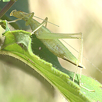
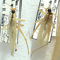 In some species of insects, the wings of the males and females
differ. In katydids and crickets, the tegmina of the males are broader and
rounder and include a stridulatory area consisting of a file on one one wing
and scraper on the other. These are rubbed together to produce sound. The
tegmina of the females lack those organs, and may be thinner and more pointed,
posteriorly, than those of the males. In this photograph, a female tree
cricket is on the left, and a male on the right.
In some species of insects, the wings of the males and females
differ. In katydids and crickets, the tegmina of the males are broader and
rounder and include a stridulatory area consisting of a file on one one wing
and scraper on the other. These are rubbed together to produce sound. The
tegmina of the females lack those organs, and may be thinner and more pointed,
posteriorly, than those of the males. In this photograph, a female tree
cricket is on the left, and a male on the right.
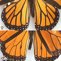 In Monarch butterflies, the males have a small scent gland,
near one of the veins on their hind wings, while the females do not have
this structure. In this photograph, the hind wings on top are from a female,
while the set of hind wings, below, is from a male.
In Monarch butterflies, the males have a small scent gland,
near one of the veins on their hind wings, while the females do not have
this structure. In this photograph, the hind wings on top are from a female,
while the set of hind wings, below, is from a male.
As time and specimens
allow, view and draw a variety of wings found on different types of (pinned)
insects, looking for as many of the types listed above as are available.
Which type(s) of wings does your grasshopper have?
Abdomen
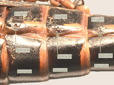 The abdomen clearly shows its segmentation. Each
segment has a sclerotized dorsal portion, here called the tergum,
(terg = back;) and a sclerotized ventral sternum
(stern = breast, breastbone), again joined by a lateral membranous
area. (Note: you will also see the words tergite and sternite used.)
The abdomen clearly shows its segmentation. Each
segment has a sclerotized dorsal portion, here called the tergum,
(terg = back;) and a sclerotized ventral sternum
(stern = breast, breastbone), again joined by a lateral membranous
area. (Note: you will also see the words tergite and sternite used.)
Spiracles (spiracl = an air hole) occur along
the sides of the abdomen, between the edges of the sternites and tergites.
They are located in small, sclerotized regions, one pair per segment.
Look
closely to see the opening of the spiracle in each segment and the lid-like
structure that controls the
aperture of the spiracle. In hot, dry weather insects can close their
spiracles to help avoid water loss (unless they need to take in more oxygen).
Observe the structure of the spiracles under the dissecting scope.
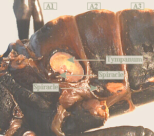 One abdominal structure that is unique to grasshoppers is the
tympanum (tympanum = a drum). In general, those types of
insects which produce sound also have some structure which allows them to
hear sound. In grasshoppers, this auditory structure consists of a pair of
tympana located on the sides of the first abdominal segment. True to their
name, these detect sound by vibrating like a drum head.
One abdominal structure that is unique to grasshoppers is the
tympanum (tympanum = a drum). In general, those types of
insects which produce sound also have some structure which allows them to
hear sound. In grasshoppers, this auditory structure consists of a pair of
tympana located on the sides of the first abdominal segment. True to their
name, these detect sound by vibrating like a drum head.
Make sure to
include the tympanum and spiracles on your grasshopper illustration.
Structures of the Posterior End:
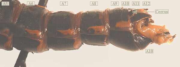
Male Lubber Grasshopper, Posterior Tip of Abdomen
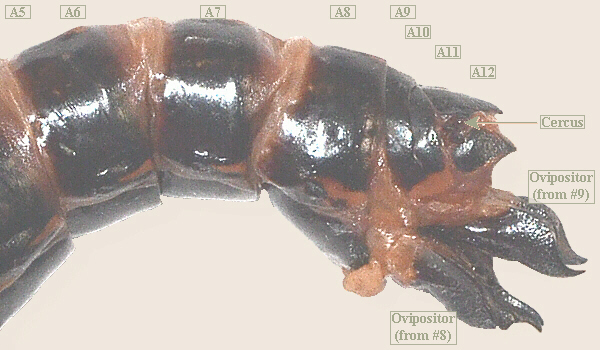
Female Lubber Grasshopper, Posterior Tip of Abdomen
Finally! Now you get to
figure out whether your grasshopper is a male or female. Examine these
photographs and compare them with your grasshopper to determine which you
have. Also, look for, draw, and label the structures listed below.
- The anus is a side-to-side slit on the last segment between
the sternite and tergite, but probably wont be very obvious (and, the
last few segments are so highly modified, it can be difficult to
differentiate among them).
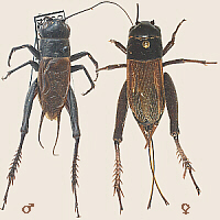
- The cercus (sing.)/cerci (pl.) (cercus = tail)
are a pair of sensory projections arising between the last two segments
(considered to be appendages of the eleventh abdominal segment). These
are sensitive to air currents, including those generated when you are
attempting to swat or catch an insect. In general, an insects nervous
system works such that when it senses an air current (= attack?), it
runs or flies away.
In this picture, the cricket on the left is a male. The two filaments
projecting from his back end are his cerci. The female has them, too,
but hers are folded in towards the center, and thus, are not visible.
- The stylus (sing.)/styli (pl.) (styl = a pillar,
stake, pointed instrument) lie between the cerci on the males
(considered to be appendages of the twelfth abdominal segment). Females do
not have styli compare. In many male orthopterans, these are modified
as claspers to grab the females genitalia during mating. These are not
easily seen in the male grasshopper, but in some insects, such as the
American Cockroach, they are quite distinct and useful in differentiation
of the sexes.
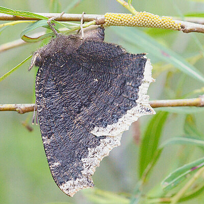
Female Mourning Cloak Laying Eggs on Willow
- The ovipositor (ovi = egg) is present on the ventral
side of the end of the abdomen in females only. Technically,
the ovipositor is derived from appendages of
abdominal segments 8 and 9.
The cricket on the right, above, is a female. When she
was alive, the long projections from her back end fit together to form
the ovipositor, serving to lay eggs in damp soil. The female
grasshopper uses the four parts of her ovipositor to dig a hole in
damp soil and inserts the rear portion of her abdomen into that
hole to lay her eggs in a cluster. This female
Mourning Cloak does not appear to have a noticeable ovipositor hers
is tiny and suited for the kind of egg-laying that she does.
- Subgenital plate This is the last abdominal tergite just
anterior to the genitalia (abdominal segment 8 or 9), and is more prominent
in some types of insects than in others. In cockroaches, for example, the
subgenital plate is noticeably different in males and females, thus useful
to differentiate the sexes.
Determine your grasshoppers sex. Draw and label pictures of the back
ends of both sexes (so you can remember whos who).
Examine and compare the ventral side of the subgenital plate of a male and
female grasshopper. How large and distinguishable is/isnt the subgenital
plate? Is there a difference between males and females?
Internal Anatomy
Longitudinal Cuts
To expose the internal structures, if not done already, first
remove the wings and legs. Then carefully make two shallow,
longitudinal (front to back) cuts through the dorsal (back) body wall of the
abdomen and thorax about ⅛ in. (3 mm) to either
side of the midline. Carefully remove the dorsal piece (save)
caution: the heart and aorta are right underneath and may stick to the
piece you are removing. Pin
the sides open onto the dissecting tray to better expose what
is inside.
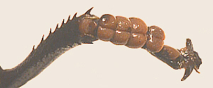
Circulatory System: Heart and Dorsal Aorta
Silkworm Heart
Silkworm Heart, Close-up
Heart and Aorta
This may come off with the top flap or it may stay on the inside. It is a
dorsal tube along the midline and lies on a thin, muscular sheet which may
come off with the tergum. Blood is pushed from posterior to anterior and
out into the head cavity. The blood flows from there back through the body,
thus this is an open circulatory system (make a sketch in your notebook of
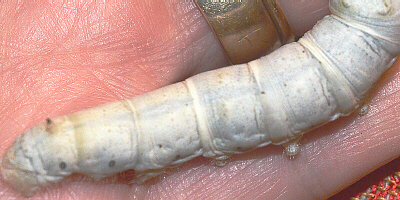
the blood flow pattern). In some insects, such as a last instar (mature)
silkworm caterpillar (examine if available), it is possible to see the heart
beating. In the silkworm photographs, above, the heart/aorta is the darker
gray line up the center of the back.
Digestive System
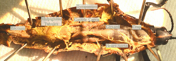
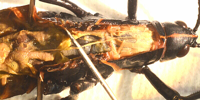
Male Lubber Grasshopper Digestive System
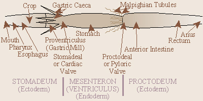
The parts are listed below from front to back. Note: the
digestive systems of many insects are somewhat- to highly-modified from this
general plan.
- Esophagus (eso = within, inward; phago = to eat)
leads from the mouth to the crop.
- Crop A food-storage area.
- Proventriculus/Gizzard For further shredding of food (beyond
what was done by the mandibles).
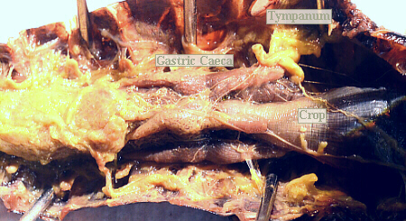
- Gastric Caeca (gastro = stomach; caec = blind)
Finger-like projections from the anterior end of the stomach in many insects.
They provide more surface area for digestion.
- Stomach Enzymes for digestion of food.
- Malpighian Tubules These are the fine yellowish threads in the
region where the stomach and intestine join. They serve an excretory
function and void the excrement into the intestine where it is passed out
along with the feces.
Of interest is the excretion of a group of shiny blue
or green flies called blow flies. In these insects, the excreted material
is a chemical called allantoin, known to be a cell-proliferant - a
substance which promotes healing. The maggots (aseptically-reared) of these
flies are used to treat deep, gangrenous wounds because they will only eat
the dead tissue and not harm the live tissue, plus the allantoin they
excrete promotes healing of the wound. Wounds treated with maggot therapy
heal faster and with less scarring than untreated- or
conventionally-treated wounds.
- Intestine Behind the stomach. Further digestion and
absorption.
- Rectum The posterior portion of the intestine leading to the
anus.
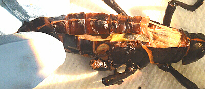
Reproductive System
- Paired Gonads are above the posterior portion of the digestive
tract.
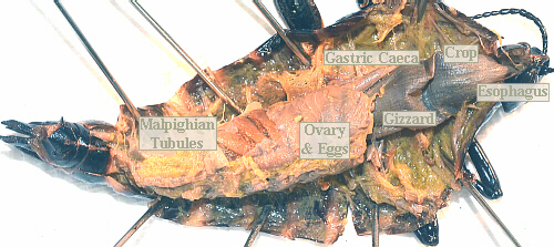
- Female System Ovary (ova, ovi = egg) walls are
often very thin, virtually invisible. Extending posteriorly from the
ovaries are the oviducts which pass around the sides
of the intestine and fuse beneath to form the vagina.
In most insects there is a small
pouch, the seminal receptacle, off the vagina where sperm are stored
and nourished and from which they go to fertilize the eggs as the eggs pass.
A number of insects such as queen bees and ants only mate once in their
lives, thus must store sperm for future use.
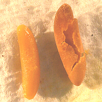
- In females where ovaries are distended with eggs, these
occupy a large space, as in the photo above. You may be able to dissect
open an egg to observe whats inside, as in the photo to the left,
although I believe they may be fertilized as they are laid, and do most, if
not all, of their development after they are laid. Thus, like this egg,
they probably will be undifferentiated will probably not contain a
recognizable embryo. That is not true in all species of insects. Some
cockroaches, for example, hold their eggs internally until they are ready
to hatch, and in that case, a dissected egg might yield an embryo.
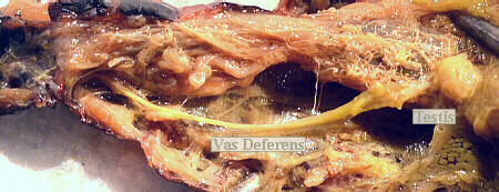
- Male System Testes (note: testis is singular and
testes is plural) appear as a translucent, whitish cluster of
finger-shaped lobes on either
side, imbedded in the mass of whiter-colored fatty tissue. Vasa
deferentia lead from the testes and join below the intestine to form the
single ejaculatory duct (e- = out, from; jacul = throw).
Males of some insects (for example, scarab beetles) have a sclerotized
intromittent organ, the aedeagus (aedeag = the genitals).
Males of some insects have the cerci and/or
styli modified as
claspers to hold the female. In most insects, sperm are transferred in a
spermatophore (phora = to bear, carry a structure which is
attached to the females genitalia and from which the sperm enter her body).
Compare your grasshopper with one of the opposite sex being dissected by
your classmates to observe the reproductive systems of both sexes.
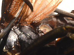
Respiratory System
 This is a fine network of tracheal tubes bring oxygen
directly to the
tissues, thus in most insects, the blood does not function in carrying
oxygen and has no hemoglobin (some have hemocyanin which is also based on
porphyrin, but with copper in the center, and that gives their blood a blue
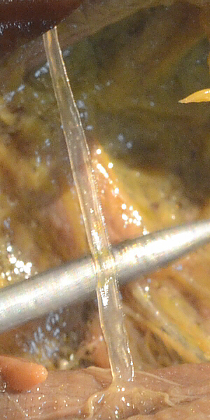
or greenish color). The respiratory system appears as
silvery-white threads of various diameters.
As in the first photo, small tracheal tubes extend from the spiracles
into the body and unite with one of the two (left and right) longitudinal
tracheal trunks that extend the length of the body (Also in the photo,
the clear,
port-hole-looking structure is the inside of the tympanum.).
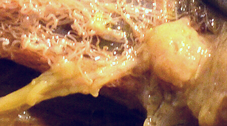
This is a fine network of tracheal tubes bring oxygen
directly to the
tissues, thus in most insects, the blood does not function in carrying
oxygen and has no hemoglobin (some have hemocyanin which is also based on
porphyrin, but with copper in the center, and that gives their blood a blue
or greenish color). The respiratory system appears as
silvery-white threads of various diameters.
As in the first photo, small tracheal tubes extend from the spiracles
into the body and unite with one of the two (left and right) longitudinal
tracheal trunks that extend the length of the body (Also in the photo,
the clear,
port-hole-looking structure is the inside of the tympanum.).
The second
photo is a close-up view of one of the incoming trachea tubes. Notice
the barely-visible rings around that tracheal tube to support it,
similar in function to the rings that support our trachea and help to keep
it open.
From the longiudinal tracheal trunks, many smaller tracheal tubes branch
off to deliver air to all the body organs.
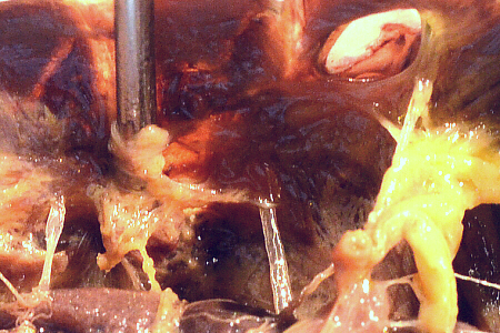
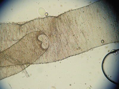 As in this third photo, make a wet mount of and observe a portion of a
tracheal tube under the microscope to see the thickened rings of tissue
(analogous to the rings you can feel in your own throat or some vacuum
cleaner hoses) which strengthen it.
As in this third photo, make a wet mount of and observe a portion of a
tracheal tube under the microscope to see the thickened rings of tissue
(analogous to the rings you can feel in your own throat or some vacuum
cleaner hoses) which strengthen it.
Muscular System
 All muscles lie within the exoskeleton and attach to it to
pull it in some direction (as in our bodies, muscles can only contract and
pull muscles cannot push). Muscle tissue is probably most visible in the
thorax where it
controls walking and flight. Interestingly, there are no muscles in
insect wings. Flight is controlled by changing the shape of the thorax,
the sclerotized sides of which act like fulcrum points under the wings.
Vertical muscles pull the notum down, so the wings, stretched across the
fulcrum points, go up like a teeter-totter. Then, longitudinal muscles pull the segment together,
front-to-back, which causes the notum to buckle upwards, and that, in turn,
causes the wings, still stretched across the fulcrum points, to go down.
There are several other sets of muscles involved in angling the wings for
lift and steering, and the actual path of the wings is a figure-eight.
Many insect movements are also brought about by hydraulic pressure: by
forcing blood/body fluids into certain areas, movement is brought
about.
All muscles lie within the exoskeleton and attach to it to
pull it in some direction (as in our bodies, muscles can only contract and
pull muscles cannot push). Muscle tissue is probably most visible in the
thorax where it
controls walking and flight. Interestingly, there are no muscles in
insect wings. Flight is controlled by changing the shape of the thorax,
the sclerotized sides of which act like fulcrum points under the wings.
Vertical muscles pull the notum down, so the wings, stretched across the
fulcrum points, go up like a teeter-totter. Then, longitudinal muscles pull the segment together,
front-to-back, which causes the notum to buckle upwards, and that, in turn,
causes the wings, still stretched across the fulcrum points, to go down.
There are several other sets of muscles involved in angling the wings for
lift and steering, and the actual path of the wings is a figure-eight.
Many insect movements are also brought about by hydraulic pressure: by
forcing blood/body fluids into certain areas, movement is brought
about.
Nervous System
Carefully remove
(or pull to one side) the digestive, reproductive, and respiratory
systems. Under them, you should see a thin muscle sheet. Gently
remove that muscle sheet. Examine, draw, and label pictures of the nervous
system.
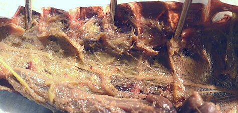
As in the photo below, the nervous system (NS) lies on the
ventral surface of
the body covered by that thin muscle sheet. The NS is a yellowish-white,
double, ventral cord running between small ganglia of nerves
in each segment. This NS is very primitive with each segmental
ganglion exercising much control over its segment. As can be seen in
this photo (and hopefully, in your grasshopper), the ventral nerve cord
actually consists of two parallel nerve cords.
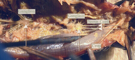
Notice, as in the following photo, that the thoracic ganglia
are larger than those in the abdomen. Why do you think this is? Notice that
anterior to the last large thoracic ganglion, the two strands/halves of
the nerve cord split and separately continue anteriorly.
Also, to the sides of the nerve cord, where the nerve cord
starts to split, in the anterior portion of the thorax, there are grape-like
clusters of small, rounded, knobby structures that are the salivary glands.
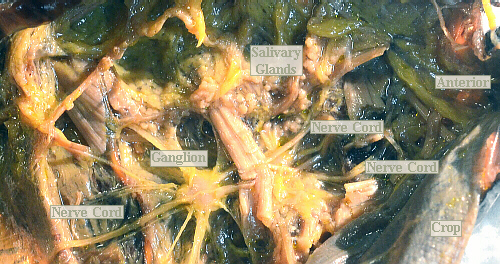
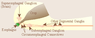
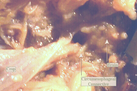
In the lower head, there is a subesophageal ganglion. Then
the double cord splits and goes around either side of the esophagus, meeting
at the supraesophageal ganglion (brain) which controls eyes, antennae,
etc. In the following photo (and its enlargement, below), one of the circumesophageal connectives is
barely visible.
Note that whereas the vertebrate NS is dorsal and a hollow
infolding of
ectoderm tissue, the arthropod NS is ventral, solid, and mesodermal in origin.
Although most insect behavior is instinctive, some learning ability is
present. Supposedly, roaches can be taught to run a simple Y maze.
Other Things to Include in Your Notebook
Make sure you have all of the following in your lab notebook:
- all handout pages (in separate protocol book)
- all notes you take as you read through the Web page and/or
during the introductory mini-lecture
- all notes and data you gather as you perform the lab
- labeled drawing(s) (yours!) of
- overall external anatomy (dorsal and/or side view)
- mouthparts
- legs
- ends of male and female abdomens
- internal anatomy
- digestive system
- nervous system and brain
- any other insect mouthparts, legs, wings, etc., that were
examined and compared
- answers to all discussion questions, a summary/conclusion in your
own words, and any suggestions you may have
- any returned, graded pop quiz
Based on printed protocol, background, artwork, and additional information
Copyright © 1990, photo of male roach Copyright © 2002,
modification for grasshopper and new photos Copyright © 2012, 2013
by J. Stein Carter. All rights reserved.
Photo of female roach and babies Copyright © 2001, and
Chickadee photograph Copyright © by David B. Fankhauser
This page has been accessed  times since 25 Dec 2012.