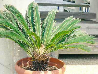
Click on This Sago Palm for More Cycad Photos
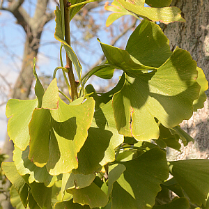
Ginkgo Leaves
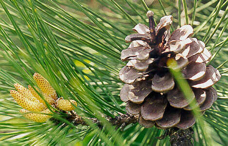
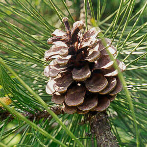
Male and Female Pine Cones (Click for More Photos)
As land-dwelling plants developed further past ferns, several modifications were developed for water conservation. These included:
In the gymnosperms, the spores produced by the sporophytes (phyto = plant) are of two different sizes/functions. Microspores (micro = small) give rise to the male gametophyte and megaspores (mega = large) to the female. The gametophytes are small and dependent on the sporophytes, as are the newly-formed embryo sporophytes. The male gametophyte, also known as pollen (poll, pollen = fine flour), is carried, often by wind, to the vicinity of the female gametophyte, and the sperm that it produces is not dependent on free surface water to reach the egg.
The gymnosperms include such plants as pines, cedars, spruces, yew, Ginkgo trees, and cycads. Much of our lumber and paper pulp comes from gymnosperms. The largest and oldest living organisms, the sequoias, are also gymnosperms and may reach heights of over 300 ft. and ages of at least 4000 years.
Pine is the most commonly studied example of a gymnosperm because of its ubiquitous distribution.
Examine, take notes on, and/or draw the various material indicated below as available and as time allows. Especially note which are the 2n sporophyte or 1n gametophyte generation. In subsequent field hikes, we should be able to examine pine trees with male and female cones, and ginkgo trees. If/when you are at a store that sells houseplants, check to see if they have Sago Palm (a cycad) for sale (typically as a bonsai), and observe it to see what it looks and feels like.
Pine is the most commonly-studied example of a gymnosperm.
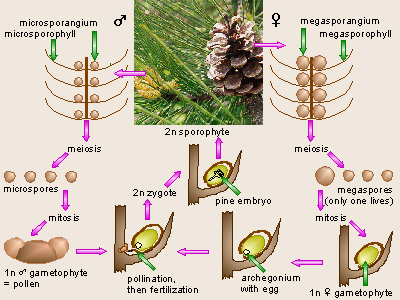
Pine Life Cycle
Draw the life cycle of pine and indicate which structures/generations are 1n or 2n. Make sure you understand this life cycle.
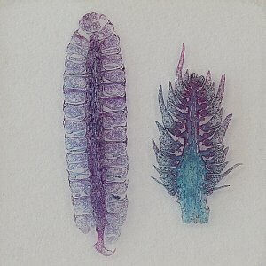
Prepared Slide of Male and Female Pine Cones
In pine as in other vascular plants, the sporophyte (the
spore-producing generation), or pine tree is the
dominant generation. It consists of an underground network of roots
supporting an upright stem (the tree trunk), which bears whorls of branches.
If the lower branches are shaded, as in a forest situation, they frequently
drop off, hence plants growing alone in the middle of a lawn may appear
quite different from those of the same species growing in a woods.
The leaves, called needles, typically occur in clusters of two
or more. The
sporophyte bears two types of reproductive structures referred to as male
and female cones (Technically these are not true male and female structures
because they do not
directly produce gametes, but they are are designated male or female
based on the sex of the gametophytes which develop from the spores which they
produce). Each cone is considered to be a modified branch
with a number of modified leaves, called scales or sporophylls.
Each sporophyll bears a structure called a sporangium in which the
spores are produced. Unlike the ferns, in gymnosperms the spores as well as
the gametes come in two sizes: separate microspores (produced in
microsporangia, and which develop into male gametophytes) and
megaspores (produced in megasporangia, and which develop into
female gametophytes) (micro = small; mega = large, great).
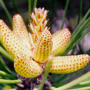
Young Male Pine Cones

Older Male Pine Cones
The male (staminate) cones typically are found in clusters at the tips of lower,
side (lateral) branches, and usually take several years to develop. In these
cones, the modified leaves are called microsporophylls (phyll
= leaf). Each microsporophyll bears two microsporangia within which
the microspores are produced. Within the
microsporangia, the microspore mother cells undergo meiosis to form
four haploid microspores, the start of the male gametophyte generation (the
pollen note, pollen is not the same thing as sperm, rather it is a
gametophyte).
Still inside the microsporangium, each microspore divides
and grows to form a four-celled (four nuclei, anyway) male
gametophyte, also known as pollen which contains
two sperm nuclei. A grain of pine pollen also has two large air sacs to
make it buoyant in the wind, and these give the pollen a Mickey Mouse
hat appearance.
If available, examine live male (staminate) cones. Examine (and draw and label) the male cone from the prepared slide of a longitudinal section (l.s.) of male and female pine cones (Carolina #B500a). Pay attention to the overall appearance, but pay closer attention to one of the microsporophylls and its contents. Thus, also identify and draw microspores and pine gametophytes (pollen).
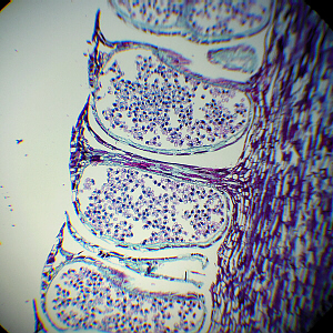
Male Pine Cone, l.s.
Pine Pollen in Microsporangium
Soon after meiosis, each microspore undergoes two successive
mitotic divisions to form a four-celled male gametophyte or
pollen grain. These four cells include two, small, flat prothallial
cells, a large tube cell, and a generative cell (which will form the sperm
nuclei).
Pollination is the transfer of the whole male gametophyte
to the female plant. In pine, the pollen is blown by wind. Note that
this is NOT the equivalent of fertilization that must still occur
later!
If you did not already do so, on the same prepared slide, examine the pollen grains within your male cone. If live male cones are available, you could, optionally, make a wet mount of some pollen from them. Often all four cells, especially the prothallial cells are difficult to see. Note also the pair of air sacs or wings which enable the pollen to be wind-disseminated (and give it a Mickey Mouse hat appearance).
Female Pine Cone
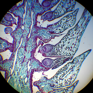
Female Pine Cone, l.s.
The female cones typically form higher up in the tree, and also usually
take several years to develop. These cones consist of a number of scales
which many botanists consider to be megasporophylls (leaves) while
others consider them to be modified branches.
In a slide of a longitudinal
section of a young cone, a small bract can often be seen attached to
the underside of each scale. On the upper surface of each scale
is a side-by-side pair of ovules, only one of which will be visible
on the slide. The central area of the ovule is called the nucellus
(the megasporangium) which contains the megaspore mother cell.
Surrounding the nucellus is an integument. Near the base
of the scale there is a gap in the integument called the micropyle
(pyle = gate, orifice) where the pollen grains enter.
Each megaspore stays within its megasporangium and goes through several
mitotic divisions to become a female gametophyte, which then develops an
archegonium with an egg in it.
Below follow several photographs of megaspores and female
gametophytes in various stages of development.
Examine (and draw and label) the female cone from the prepared slide of a longitudinal section (l.s.) of male and female pine cones (Carolina #B500a). There may be pollen grains within the micropyle on your slide (as shown in the last photo, below), or if your section is through the side of the ovule, the micropyle may not be obvious. If live female cones are available, examine them to view the side-by-side ovules or ovule scars on each of the scales.
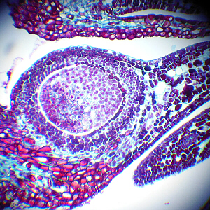
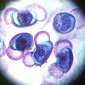
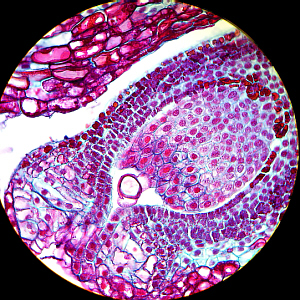
Pine Megaspore/Female Gametophyte

Pine Megaspore/Female Gametophyte
Pine Megaspore/Female Gametophyte
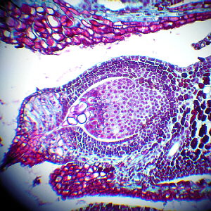
Pine Megaspore/Female Gametophyte
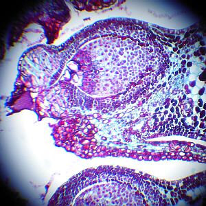
Pine Megaspore/Female Gametophyte
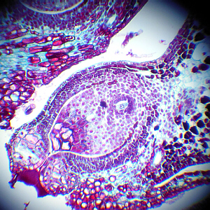
Pine Megaspore with Pollen Grain Past Micropyle
When a pollen grain lodges between the stem of the female cone and a megasporophyll/ovule (or passes through the micropyle, as shown in the last photo, above), we say that pollination has occurred. Then, to accomplish fertilization, once the pollen grain has entered the micropyle, a tube from the pollen (called a pollen tube) germinates and grows through the nucellus. This stimulates the megaspore mother cell to undergo meiosis, producing four megaspores, only one of which in each ovule develops/grows into a female gametophyte. The female gametophyte eventually is multicellular. Most of it is undifferentiated tissue which will be used to store nutrients, but near the micropyle, several archegonia (archae, archeo = ancient, first, beginning; goni = seed; -ium = small; gonium = production) are formed, each containing an egg.
Examine the female pine cone on your prepared slide to see if you can identify any of these structures.
As mentioned above, pollination is the transfer of pollen to the female cone, and is not the same as fertilization. After a pollen grain enters the micropyle and contacts the nucellus of the female plant, as mentioned, a tube from the pollen grain begins to grow through the nucellus. The nucleus of the tube cell remains near the tip of this tube, apparently directing its growth. Within the pollen, the generative cell divides to form a stalk cell and a body cell. The nucleus of the body cell divides once more to form two sperm nuclei. When the pollen tube penetrates the egg, the sperm nuclei travel down the pollen tube, then enter the egg. One of them unites with the egg nucleus (fertilization) and the other disintegrates. Even though several archegonia are formed, only one egg is fertilized and only one zygote develops per ovule. The other sperm nucleus and all other eggs and archegonia in that female gametophyte disintegrate so one zygote per ovule is left.
Make sure you understand the difference between pollination and fertilization.
As the cells of the new zygote divide, an embryo sporophyte (2n) is formed in the midst of the female gametophyte (1n). The female gametophyte (1n) stores up food, especially oils and proteins, for the embryo to use until it sprouts and begins to do photosynthesis. The nucellus (2n) becomes a thin, papery layer and the integument (2n) becomes a hard seed coat. A portion of the scale (2n) separates from the scale to form a wing for the seed. The embryo within the female gametophyte has definite parts, visible in a cross section of the seed. The central axis of the embryo is called the hypocotyl (hypo = under, beneath, cotyl = cup, cavity, socket) because it is located below the cotyledons. The bottom end (near the micropyle) is called the radicle (radix = root) and will become the root. At the opposite (top) end is a cluster of finger-like leaves called cotyledons. In the center of them is a mound of tissue called the epicotyl (epi = upon, over, beside) which, technically is above them, and will become the new stem (trunk) and leaves (needles).
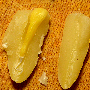
Pine Seed and Embryo
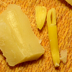
Pine Seed and Embryo
When the seed is mature or ripe, it separates from the female cone and is carried by the wind to a new location. When the seed germinates, the embryo continues to grow into a new pine tree. Initially, it uses the nutrients provided by the female gametophyte until its first leaves are above ground and large enough to do photosynthesis.
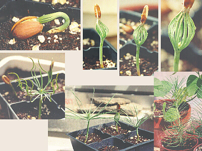
Pine Seeds Sprouting
Examine a pine nut carefully sliced in half lengthwise to view the embryo. Optionally, in the lab kitchen or at home, taste a pine nut. These are used in the cuisine of several cultures, with the most notable use being in pesto sauce. If whole pine seeds are available (now or later, while on a hike), notice how the wing on the seed helps it to be blown/dispersed by the wind.
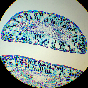
Pine Needles, x.s.
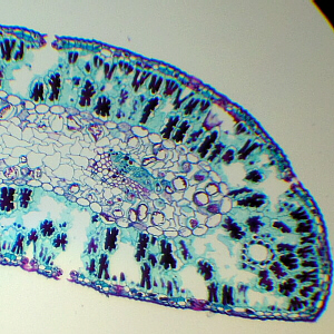
Pine Needle, x.s., Close-up
A pine needle is a modified leaf. It is very compact to prevent
water loss through evaporation.
Time does not allow us to view this slide of a cross-section of a pine needle.
It contains many of the same tissues that Angiosperm leaves do,
including epidermis (derm = skin), stomates
(stoma = mouth), palisade cells, spongy mesophyll
(meso = middle, phyll = leaf), and vascular bundles
(veins), but they are arranged slightly differently to accomodate the
rounded shape of the pine needle.
(Click for more photos of gymnosperms.)
(serves 24 people)
Mix it all in the blender until it turns into a paste. Serve over (cooked) whole grain spaghetti.
Make sure you have all of the following in your lab notebook: