Earthworm Anatomy
Background Phylum Annelida:
(annel = a little ring, a ring)
Annelid classes include
- Class Oligochaeta (oligo = few, scant; chaeta, setum
= bristle), earthworms with few setae;
- Class Polychaeta (poly = many), marine worms with
projections from each segment (parapods para = beside,
near; poda = foot) which serve as gills and bear many setae;
- Class Hirudinea, leeches.
Annelids are segmented, both inside and out. They have a
tube-within-a-tube body plan, a closed circulatory system with five pairs of
hearts, and a ventral, solid, nervous system which arises from mesoderm
tissue. (Compare this with the vertebrate nervous system which is dorsal,
hollow, and of ectodermal origin.). Many of the internal organs, for
example, the paired nephridia (nephri = kidney) which serve an
excretory function analogous to our kidneys, are repeated in each segment.
Annelids have both longitudinal and circular muscles, so have better control
over their movement than the Nematodes (which only have longitudinal
muscles) do.
Leeches are famous for their diet: they suck blood. Some have sharp jaws
to slit their hosts skin, while others secrete an enzyme to digest a hole
in the victims skin. It is to the leechs advantage if the would-be victim
remains unaware of the leechs presence, thus most leeches secrete some kind
of anesthetic (an = not, without; aesthet = sensitive,
perception) so the host does not feel their attack. To prevent the blood
from clotting before they ingest it, leeches also secrete an anticoagulant
(anti = against, opposite; co- = with, together;
ageve = to move, put in motion; coagulum = rennet). For years,
people thought disease was caused by too much blood, so the Medicinal Leech
Hirudo medicinalis was used to suck some out. Typically a medicinal
leech, once attached, may suck for a couple hours, perhaps ingesting between
one to two ounces of blood, many times its own body weight. While we now
have different theories as to the cause of diseases, medicinal leeches still
have uses in modern medicine! When someone severs a finger which must be
surgically reattached, since all the capillaries are dysfunctional, blood
flowing into the finger via the reconstructed arteries has nowhere to go but
into the tissue. This leads to problems with edema in the area. Also,
because of this poor circulation, the reattached tissues cannot get the air
and nutrients they need to heal properly. It has been discovered that
letting a leech suck on the end of the reattached finger will a) help reduce
edema, and b) create a sort-of blood flow that will allow nutrients and air
to get to the reattached tissues so they heal better and more quickly.
Additionally, the anticoagulant secreted by the Medicinal Leech is very
powerful, and a leech bite (in a person whose blood usually clots normally)
can take a couple days to stop bleeding and form a scab. Research is being
done on using this powerful anticoagulant to help heart attack and stroke
victims whose problems are caused by blood clots.
Marine worms have a pair of gill-like structures called
parapods projecting from the sides of each segment. It is believed
that the early annelid ancestors of insects (and other arthropods) may have
looked like this (and the parapods later became modified into true legs).
Background Earthworms:
In this lab, you will become familiar with the external and
internal anatomy of the earthworm, Lumbricus terrestris.
As members of Phylum Annelida, earthworms have bilateral
(bi = two, later = the side) symmetry and a tube-within-a-tube
body plan. A distinguishing feature of this phylum is the division of the
body into segments. This segmentation is both external and internal
with many structures/organs repeated in each segment. The body has three
tissue layers:
- ectoderm (ecto = outside, out, outer; derm =
skin)
- mesoderm (meso = middle)
- endoderm (endo = within, inner)
The coelom (coel = hollow) is a cavity between
the layers of the mesoderm. The circulatory system is a closed
circulatory system, meaning that the blood remains within the blood
vessels (in an open system, the blood bathes the body organs for at least
part of its journey).
Earthworms live in the soil, working their way through it to
ingest and digest organic matter within the soil. They play an important
part in aerating and fertilizing the soil.
Earthworms exchange air (breathe) through their moist skin,
thus if a worm dries out, besides the dangers of dehydration, it cant get
air so it dies. However, earthworms also cannot live underwater, and if the
soil in which they are living becomes totally filled with water during a
heavy rain, they will all come to the surface so they dont drown (and end
up on the sidewalk where they subsequently dry out after the rain stops).
Earthworms are hermaphroditic and a pair of worms fertilize
each other.
External Anatomy:
Obtain a dissecting tray (Please note that this is not a toy.
Try to avoid unnecessary pin holes in the wax and DO NOT CARVE, chop, or
otherwise mutilate the wax.) and a set of dissecting tools, as well as your
dissecting scope and its light. (Eventually, you will also need your
regular microscope.) Also, obtain a rinsed, preserved earthworm.
As you do your dissection, compare what youre seeing in your worm with
the large worm model. Draw a labeled illustration of and take notes on
everything you view.
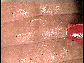
External View, Four Pairs of Setae
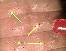
Labeled Setae
- Feel the bristly setae
(seta = bristle) on each segment, along the sides and bottom. Note
that there are four pairs. Two pairs are on the sides (laterally
later = the side) and two pairs are on the bottom (ventrally
venter = the underside, belly).
- Note the conspicuous swelling near
the anterior (the front ante = before) end called the
clitellum (clitell = a pack saddle). The smooth side (often
darker) is the top (dorsal dorso = the back) and the
segmented side (often lighter) is the bottom (ventral).
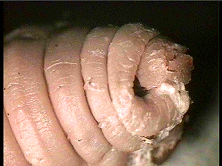
External View, Mouth
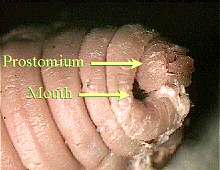
Labeled Mouth
- On the very anterior tip of the worm
is a projection called the prostomium (pro = before, in front
of; stoma = mouth). The mouth lies ventrally between this and
the first segment. Count the number of segments between the front of the
worm and the front edge of the clitellum, as well as the number of segments
included in the clitellum.
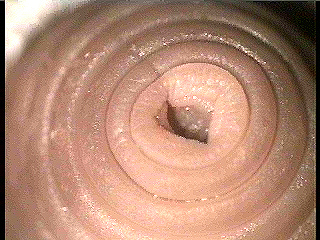
External View, Anus
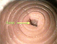
Labeled Anus
- On the posterior (the rear
post = behind, after) end, note the slit-like anus in the last
segment.
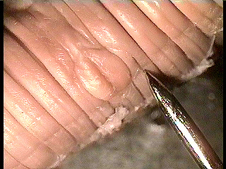
External View, Genital Openings
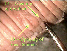
Labeled External Genitalia
- On the ventral (bottom)
surface of segment 15, note the pair of (larger) openings of the vasa
deferentia. On the ventral surface of segment 14, you can see the
(smaller) openings of the oviducts (vasa = a vessel, duct;
deferens = to carry away; ovi = egg).
Internal Anatomy:
- On the dorsal surface of the
earthworm, beginning at the clitellum, cut a slit posteriorly (toward the
rear) for about 25 segments. You must cut very shallowly to avoid
cutting the internal organs. Turn the scissors and cut anteriorly to the
prostomium, again, being careful not to cut the internal organs.
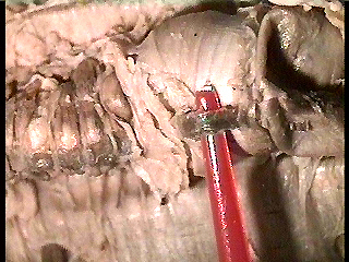
Internal View, Dorsal Blood Vessel
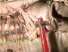
Labeled Dorsal Blood Vessel
- Pin the worm to the tray near the
anterior and posterior ends and every five segments (5, 10, 15, etc.) along
the body. You may have to carefully remove/cut the septa
(septum = a fence) separating the segments to open up the body wall.
The dorsal blood vessel and digestive tract should be exposed
at this point.
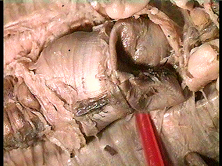
Internal View, Crop and Gizzard
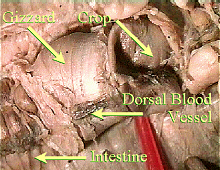
Labeled Crop and Gizzard
- Examine the digestive tract.
From the mouth back, locate the pharynx, esophagus
(eso = within, inward; phago = to eat), crop,
gizzard, and intestine. Refer to these photos, and the
large earthworm model.
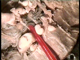
Internal View, Five Pairs of Hearts
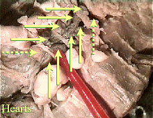
Labeled Hearts
- Examine the circulatory system.
Locate the dorsal blood vessel, smaller segmental vessels coming
from it, 5 pairs of hearts in segments 7-10 (count from the placement
of the pins), ventral blood vessel (you may have to carefully snip a
piece of intestine and hold it up to see this).
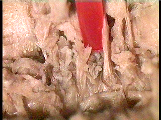
Internal View, Nephridia
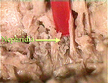
Labeled Nephridia
- To the sides/under the digestive
tract are the paired nephridia (nephr = kidney), which may be
too small to see well with the unaided eye.
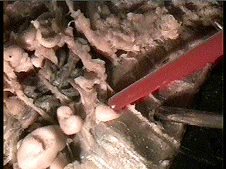
Internal View, Seminal Vesicles and Receptacles
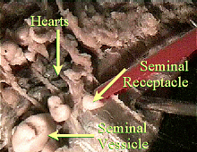
Labeled Seminal Vesicles/Receptacles
- The reproductive systems are
located under the digestive tract in approximately segments 10-15.
Earthworms are hermaphroditic (Hermes = messenger god
Mercury; Aphrodite = goddess of love Venus), that is, they have
both sexes and when they mate, they fertilize each other. Seminal
vesicles (male organs) are large, floppy, whitish structures in segments
9-13. The tubes from them to segment 15 are the vasa deferentia.
Attached to the anterior septum of segment 13 is a pair of whitish,
grape-clustered ovaries (female organs) which are very difficult to
see. The seminal receptacles, sperm-storage areas within the female
reproductive tract, are smaller, whitish organs near the seminal vesicles.
Oviducts start at the ovaries, go past the seminal receptacles, then
to segment 14, from which they open to the outside.
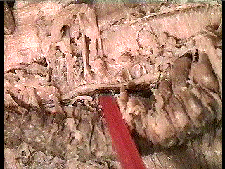
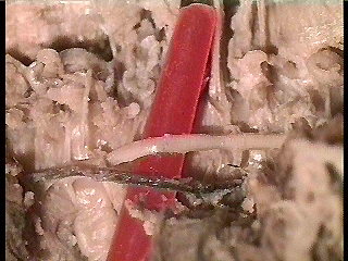
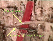
External View, Ventral Nerve Cord and Ventral Blood Vessel
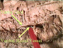
Labeled Nerve Cord & Blood Vessel
- Note the ventral nerve cord
with segmental ganglia (ganglion = a knot on a string). If
you lift a section of the digestive tract/blood vessels, the nerve cord
should be seen lying on the ventral surface of the body cavity.
Internal View, Ventral Nerve Cord and Blood Vessel
Labeled Nerve Cord & Blood Vessel
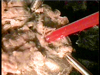
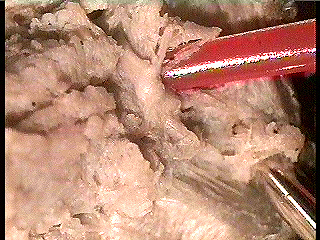
Internal View, Brain
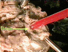
Labeled Brain
- If you look carefully at the nerve
cord, you may be able to see that it is actually two, paired cords. As you
proceed anteriorly, the last segmental ganglion (the subpharyngeal
ganglion) will be found in segment #4. From there, the two halves of
the nerve cord (the circumpharyngeal connectives) split and go around
either side of the pharynx. Above the pharynx, they come together, again,
to form the suprapharyngeal ganglion, also called the brain.
To aid you in finding your worms brain, Dr. Fankhauser has described the
worm brain as looking like the top half of a womans bikini bathing suit.
Internal View, Brain
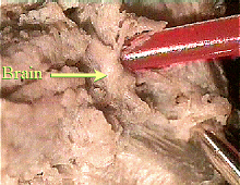
Labeled Brain
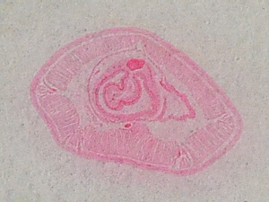
Cross-Section of Earthworm Segment
Cut a thin cross-section through your worms intestinal area
and view it with a dissecting scope. Also, view the prepared slide of
an earthworm cross section (Carolina #Z1250), and examine the large model
worm.
Cross Section, Whole Worm
Labeled Worm Cross Section
- Note/draw the layers in the body wall
from the outside in: cuticle secreted by the epidermis,
epidermis (epi = upon, over, beside) made of ectoderm
tissue, then circular muscle layer, longitudinal muscle layer,
and parietal peritoneum (pariet = a wall; peri = around;
ton = something stretched; -eum = a place where;
peritoneum = the membrane around the intestines) all mesoderm
tissues (meso = middle). Note the four areas where the setae are
located, although you may not see the actual setae
on your slide.
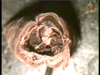
Intestinal Area, x.s., Actual View
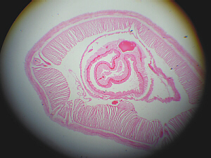
Intestinal Area, x.s., Microscopic View
- Note the coelom (coelo
= hollow), the body cavity. Often there is a tangled network visible within
the coelom which is part of the nephridia, the excretory organs.
Each nephridium opens to the outside via a nephridiopore (which may
not be visible on your slide).
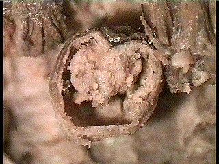
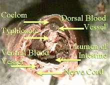
Cross Section, Central Tube
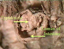
Labeled Intestinal Cross Section
- Examine the central tube. Note that
its upper (dorsal) surface is folded into the digestive tract. This portion
is called the typhlosole (typhlo = blind; solen =
channel, pipe), and helps to increase the surface area of the digestive
tract. (Note: if you carefully open a length of your worms intestine
(and clean out the soil/food contained therein), you may be able to see the
typhlosole running the length of the dorsal surface and somewhat resembling
a tightly-coiled spring.) The layers of the central tube, from outside in,
are the chloragen cells,
then thin muscular layers (both mesoderm), then the gastric
epithelium (gastro = stomach, theli = nipple), which is
endoderm tissue, as the lining of the digestive tract. The space
within the digestive tract is known as the lumen. Dorsally and
ventrally of the digestive tract run the dorsal and ventral blood
vessels. Ventrally, in the coelom, there is a nerve cord that
runs the length of the body.
Other Things to Include in Your Notebook
Make sure you have all of the following in your lab notebook:
- all handout pages (in separate protocol book)
- all notes you take as you read through the Web page and/or
during the introductory mini-lecture
- all notes and data you gather as you perform the lab
- labeled drawings (yours!) of
- overall external anatomy
- external view of anterior end
- external view of posterior end
- external, ventral view of segments 14-15
- external arrangement of setae
- overall internal anatomy (dorsal view)
- close-up of any internal organs of special interest,
such as the brain
- cross-section of intestinal segment
- answers to all discussion questions, a summary/conclusion in your
own words, and any suggestions you may have
- any returned, graded pop quiz
Copyright © 1999 by J. Stein Carter. All rights reserved.
Based on printed protocol, background, and other information
Copyright © 1989, Labeled photos Copyright © 2005 J. L. Stein Carter
Chickadee photograph Copyright © by David B. Fankhauser
This page has been accessed  times since 8 Apr 2006.