Muscles
Muscle Tissue:
There are three different types of muscle tissue.
These include the skeletal muscles which are under voluntary control
and are made of striated muscle tissue, the visceral muscles
which are under involuntary control and are made of smooth muscle
tissue, and cardiac muscle tissue which is found only in the heart.
Cardiac muscle tissue has properties in common with each of the other two
types. An interesting exception to the visceral muscles being under
involuntary control is the
diaphragm,
which controls breathing. This muscle is normally under involuntary control
(we dont usually have to think about breathing), but a person can exert a
limited amount of voluntary control also (for example, purposely holding
ones breath or breathing quickly or deeply).
Muscle cells contain filaments of two kinds of proteins,
actin and myosin, which slide past each other as the muscle
contracts. After a muscle contracts, ATP (produced in the muscle cells
mitochondria) is needed to relax the muscle and return the actin and myosin
filaments to their normal positions. When a person (or other animal) dies
and the mitochondria are no longer producing ATP, the muscles cannot relax.
This stiffening of the muscles is called
rigor mortis.
Muscle contraction is initiated when an electrical impulse from a nerve cell
reaches its associated muscle cell(s), causing positively- and
negatively-charged ions to switch places all along the muscle cell (fiber).
Movement of Ca++ ions in/out of the muscle cell (fiber) is
important in both contraction and relaxation of the muscle, so if a person
doesnt ingest enough calcium, some could be taken out of the bones to supply
the muscles with what they need to contract and relax. Any given muscle
fiber reacts in an all or none response it is either relaxed or
contracted, and the variability in contraction of the overall muscle is
based on the number of fibers which contract.
It is important to remember that muscles can only pull or
contract, not push. Thus, many muscles come in sets of antagonists
that do opposite jobs. For example, the muscle on the top of your arm
bends the arm at the elbow while the muscle under your arm straightens the
arm.
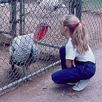
Turkey and Friend
Skeletal muscles can be further subdivided into two sub-types depending on
their use or function, and the chemical composition of each is slightly
different. Think of a Thanksgiving turkey: the first question asked is
invariably, Do you want white or dark meat? These correspond to white
and red muscle tissue, respectively. Think of where the white and
dark meat are found: dark meat is found in the legs and other constantly-used
posture muscles, while white meat is found in the breast, or flight muscles.
Turkeys and chickens arent known for flight, so these flight muscles get
infrequent, quick bursts of use. Because the posture muscles are constantly
used, they need a more constant, steady supply of oxygen. Thus red muscle
tissue contains an extra chemical called
myoglobin
which is a special protein-type molecule for oxygen storage. The presence of
myoglobin in posture muscles enables the sustained contractions necessary to
maintain proper posture and walk, so in a turkey, red muscle tissue is found
in the legs and other support muscles. The presence of myoglobin gives the
muscle tissue its red or dark color. White muscles that are only used
occasionally dont have myoglobin in their tissue. Since turkey flight
muscles are only used for short, quick flights, they dont need as much
oxygen, thus dont need myoglobin to store it, and appear white in
color. The locations of myoglobin-containing muscles in an organisms body
is a genetically-controlled,
species-specific trait. For example, some birds that spend more time flying
than turkeys and chickens also have myoglobin in their breast muscles.
Skeletal muscles can be classified as one of several different types including:
- Flexor
-
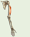
Flexor Muscle (click to show animation)
A muscle which bends a joint
- Extensor
-
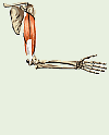
Extensor Muscle (click to show animation)
A muscle which straightens a joint
- Abductor
- A muscle which moves a body part away from the midline of the body
- Adductor
- A muscle which moves a body part toward the midline of the body
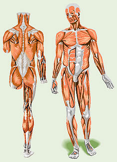
Human Muscles:
Unfortunately, your text has no good illustration of the
locations of the main muscles in the human body. The main muscles include:
- pectoralis major
- the muscles in the upper chest
- deltoid
- abductor muscle over the shoulder
- triceps brachii
- extensor muscle on the back of the arm: the term brachii
distinguishes from biceps femoris
- biceps brachii
- flexor muscle on the front of arm
- rectus abdominis
- the segmented muscles up the center front of the abdomen
- diaphragm
- internal muscle that divides the thorax and abdomen
- trapezius
- muscle that forms the back of the neck
- latissimus dorsi
- mid-back muscle over the kidneys
- gluteus maximus
- the seat or rump muscle
- sartorius
- leg-rotator muscle attached at the outer top of the thigh and inside
by the knee
- quadriceps femoris
- the main extensor muscle on the front of the femur
- gracilis
- slender muscle along the inside of the thigh, holds the legs
together
- hamstrings
- flexor muscles on the back of the thigh which include:
- biceps femoris
- the lateral or outer one: the term femoris distinguishes from
biceps brachii
- semitendinosus
- the medial or inner one
- gastrocnemius
- the calf muscle, consists of two halves and ends in. . .
- Achilles tendon
- an extension of the gastrocnemius down around the back of the
heel
Together, these two extend the foot.
Muscle Identification Practice:
Copyright © 1996 by J. Stein Carter. All rights reserved.
This page has been accessed  times since 14 Mar 2001.
(Clipart modified from Corel Presentations 8)