Reproductive System
Background Information:

Female Mourning Cloak Laying Eggs on Willow
Animals reproductive systems can be divided into the
internal reproductive organs and the external genitalia. The gonads
are the actual organs that produce the gametes. In the male,
testes (singular = testis) produce sperm, and in the female,
ovaries make eggs.
In most animals, individuals are either
definite males or definite females. However, in some species, individual
organisms are both male and female.
Hermaphroditism
is when one organism has both sexes. Earthworms and garden snails always
have both male and female organs, and when, for example, two earthworms mate,
they fertilize each other. A special variation on the theme is
sequential hermaphroditism, in which an organism changes sex during
its life. If an organism is female first and later changes to male, that
organism is
protogynous,
and if the organism is male first and changes to female, it is said to be
protandrous.
In different species, sequential hermaphroditism can be influenced by the
organisms age or size or by various environmental/climatic factors.
While most higher animals reproduce sexually, there are some
species in which the females can, under certain conditions, produce offspring
without mating.
Parthenogenesis
is the ability of an unfertilized egg to develop and hatch. This seems to be
especially prevalent among insects. Some of the giant walkingsticks at the
Zoo are females who, without mating, lay eggs that hatch into more females
generation after generation. Other insects, like some aphids, have
complicated life cycles that involve sexually-reproducing generations
alternating with parthenogenically produced generations. In honeybees,
fertilized eggs turn into females (workers and queens), while unfertilized
eggs, which are only produced in the spring, turn into males.
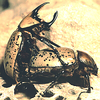
Eastern Hercules Beetles Mating
In sexual reproduction, there must be some way of getting the
sperm to the egg. Since sperm and eggs are designed to be in a watery
environment, aquatic animals can make use of the water in which they live,
but terrestrial animals must, in some way, provide the wet environment needed
for the sperm to swim to the egg. There are, thus, two major mechanisms of
fertilization. In external fertilization, used by many aquatic
invertebrates, eggs and sperm are simultaneously shed into the water, and the
sperm swim through the water to fertilze the egg. In internal
fertilization, the eggs are fertilized within the reproductive tract of
the female, and then are covered with eggshells and/or remain within the
body of the female during their development.
In species with external fertilization, at an appropriate
developmental stage, the eggs hatch, and the new young simply swim away.
However, females of species with internal fertilization must, at some point,
expel the growing young. There are three general ways of doing this:
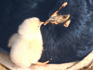
Oviparous Chicken with New Baby
- Oviparous
organisms, like chickens and turtles, lay eggs that continue to develop after
being laid, and hatch later.

Viparous Newborn Puppy
- Viviparous
organisms, like humans and kangaroos, are live-bearing. The developing young
spend proportionately more time within the females reproductive tract,
portions of which are specially-modified for this purpose. Young are later
released to survive on their own.
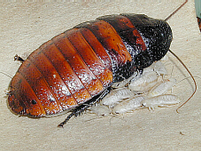
Ovoviparous Cockroach and New Babies
- Ovoviviparous
organisms, like guppies, garter snakes, and Madagascar hissing roaches, have
eggs (with shells) that hatch as they are laid, making it look like live
birth.
Human Male Reproductive System:
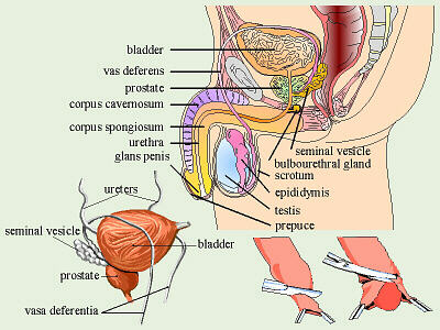
Male Reproductive System
(clipart edited from Corel Presentations 8)
The male reproductive system is illustrated to the right. Sperm are
produced in the testes located in the scrotum. Normal body
temperature is too hot thus is lethal to sperm so the testes are outside of
the abdominal cavity where the temperature is about 2° C (3.6° F) lower.
Note also that a womans body temperature is lowest around the time of
ovulation to help insure sperm live longer to reach the egg. If a man takes
too many long, very hot baths, this can reduce his sperm count. Undescended
testes (testes are supposed to descend before birth) will cause sterility
because their environment is too warm for sperm viability unless the
problem can be surgically corrected. (Undescended testes are also very
prone to developing cancer, thus if they cannot be surgically moved, they
probably will need to be removed.)
From there, sperm are transferred to the
epididymis,
coiled tubules also found within the scrotum, that store sperm and are the
site of their final maturation.
In
ejaculation,
sperm are forced up into the
vas deferens
(plural = vasa deferentia).
From the epididymis, the vas deferens goes up, around the front of, over the
top of, and behind the bladder. A vasectomy is a fairly simple,
outpatient operation that involves making a small slit in each scrotum,
cutting the vasa deferentia near where they begin, and tying off the cut ends
to prevent sperm from leaving the scrotum. Because this is a relatively
non-invasive procedure (as compared to doing the same to a womans oviducts),
this is a popular method of permanent birth control once a couple has had
all the children they desire. Couples should carefully weigh their options,
because this (and the corresponding female procedure) is not designed to be a
reversible operation.
The ends of the vasa deferentia, behind and slightly under the
bladder, are called the ejaculatory ducts. The seminal vesicles
are also located behind the bladder. Their secretions are about 60% of the
total volume of the semen (= sperm and associated fluid) and contain
mucus, amino acids, fructose as the main energy source for the sperm, and
prostaglandins to stimulate female uterine contractions to move the semen up
into the uterus. The seminal vesicles empty into the ejaculatory ducts.
The ejaculatory ducts then empty into the urethra (which, in males,
also empties the urinary bladder).
The initial segment of the urethra is surrounded by the
prostate gland
(note spelling!). The prostate is the largest of the accessory glands and
puts its secretions directly into the urethra. These secretions are alkaline
to buffer any residual urine, which tends to be acidic, and the acidity of the
womans vagina. The prostate needs a lot of zinc to function properly,
and insufficient dietary zinc (as well as other causes) can lead to
enlargement which potentially can constrict the urethra to the point of
interferring with urination. Mild cases of prostate hypertrophy can
often be treated by adding supplemental zinc to the mans diet, but severe
cases require surgical removal of portions of the prostate. This surgery,
if not done very carefully can lead to problems with urinary incontinence
or sexual performance.
The bulbourethral glands or Cowpers glands are
the third of the accessory structures. These are a small pair of glands
along the urethra below the prostate. Their fluid is secreted just before
emission of the semen, thus it is thought that this fluid may serve as a
lubricant for inserting the penis into the vagina, but because the volume
of these secretions is very small, people are not totally sure of this
function.
The urethra goes through the penis. In humans, the
penis contains three cylinders of spongy, erectile tissue. During
arousal, these become filled with blood from the arteries that supply them
and the pressure seals off the veins that drain these areas causing an
erection, which is necessary for insertion of the penis into the
womans vagina. In a number of other animals, the penis also has a bone, the
baculum, which helps to stiffen it. The head of the penis, the
glans penis, is very sensitive to stimulation. In humans, as in
other mammals, the glans is covered by the foreskin or prepuce, which
may have been removed by circumcision. Medically, circumcision is
not a necessity, but rather a cultural tradition. Males who have not been
circumcised need to keep the area between the glans and the prepuce clean so
bacteria and/or yeasts dont start to grow on accumulated secretions, etc.
there. There is some evidence that uncircumcised males who do not keep the
glans/prepuce area clean are slightly more prone to penile cancer.
Human Female Reproductive System:
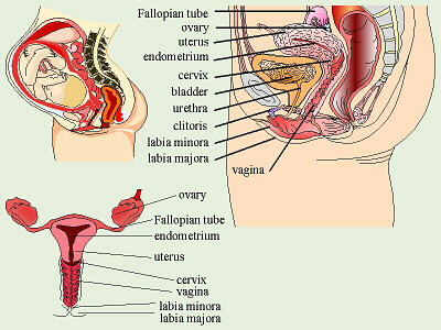
Female Reproductive System
(clipart edited from Corel Presentations 8)
The female reproductive system is illustrated to the right. Eggs are
produced in the ovaries, but remember from our discussion of
meiosis,
that these are not true eggs, yet, and will never complete meiosis and
become such unless/until first fertilized by a sperm. Within the ovary, a
follicle consists of one precursor egg cell surrounded by special
cells to nourish and protect it. A human female typically has about 400,000
follicles/potential eggs, all formed before birth. Only several
hundred of these eggs will actually ever be released during her
reproductive years. (As just an example, if a woman would ovulate from age
15 to age 55, which is a long time, thats 40 years. If wed
assume 13 28-day cycles per year, that would be 40 × 13 = 520 potential times
an egg could be released.) Normally, in humans,
after the onset of puberty, due to the stimulation of
follicle-stimulating hormone (FSH) one egg per cycle matures and is
released from its ovary. Ovulation is the release of a mature egg
due to the stimulation of leutenizing hormone (LH), which then
stimulates the remaining follicle cells to turn into a
corpus luteum
which then secretes progesterone to prepare the uterus for possible
implantation. If an egg is not fertilized and does not implant, the corpus
luteum disintegrates and when it stops producing progesterone, the lining of
the uterus breaks down and is shed.
Each egg is released into the abdominal cavity near the
opening of one of the oviducts or Fallopian tubes. Cilia in
the oviduct set up currents that draw the egg in. If sperm are present in
the oviduct (if the couple has recently had intercourse), the egg will be
fertilized near the far end of the Fallopian tube, will quickly finish
meiosis, and the embryo will start to divide and grow as it travels to the
uterus. The trip down the Fallopian tube takes about a week as the cilia in
the tube propel the unfertilized egg or the embryo down to the uterus.
At this point, if she had intercourse near the time of ovulation, the woman
has no idea whether an unfertilized egg or a new baby is travelling down
that tube. During this time, progesterone secreted by the corpus luteum has
been stimulating the endometrium, the lining of the uterus, to thicken
in preparation for possible implantation, and when a growing embryo finally
reaches the uterus, it will implant in this nutritious environment and begin
to secrete its own hormones to maintain the endometrium. If the egg was
not fertilized, it dies and disintegrates, and as the corpus luteum also
disintegrates, its progesterone production falls, and the unneeded, built-up
endometrium is shed.
The uterus has thick, muscular walls and is very small.
In a
nulliparous
woman, the uterus is only about 7 cm long by 4 to 5 cm wide, but it can
expand to hold a 4 kg baby. The lining of the uterus is called the
endometrium,
and has a rich capillary supply to bring food to any embryo that might
implant there.
The bottom end of the uterus is called the
cervix.
The cervix secretes mucus, the consistency of which varies with the stages
in her menstrual cycle. At ovulation, this cervical mucus is clear,
runny, and conducive to sperm. Post-ovulation, the mucus gets thick and
pasty to block sperm. Enough of this mucus is produced that it is possible
for a woman to touch a finger to the opening of her vagina and obtain some
of it. If she does this on a daily basis, she can use the information thus
gained, along with daily temperature records, to tell where in her cycle she
is. If a woman becomes pregnant, the cervical mucus forms a plug to seal off
the uterus and protect the developing baby, and any medical procedure
which involves removal of that plug carries the risk of introducing pathogens
into the nearly-sterile uterine environment.
The vagina is a relatively-thin-walled chamber. It
servs as a repository for sperm (it is where the penis is inserted), and
also serves as the birth canal. Note that, unlike the male, the female has
separate opening for the urinary tract and reproductive system. These
openings are covered externally by two sets of skin folds. The thinner,
inner folds are the
labia minora
and the thicker, outer ones are the labia majora. The labia minora
contain erectile tissue like that in the penis, thus change shape when the
woman is sexually aroused. The opening around the genital area is called
the vestibule. There is a membrane called the
hymen
that partially covers the opening of the vagina. This is torn by the womans
first sexual intercourse (or sometimes other causes like injury or some kinds
of vigorous physical activity). In women, the openings of the vagina and
urethra are susceptible to bacterial infections if fecal bacteria are wiped
towards them. Thus, while parents who are toilet-training a toddler usually
wipe her from back to front, thus imprinting that sensation as feeling
right to her, it is important, rather, that that little girls be taught to
wipe themselves from the front to the back to help prevent vaginal and
bladder infections. Older girls and women who were taught the wrong way
need to make a conscious effort to change their habits.
At the anterior end of the labia, under the pubic bone, is the
clitoris,
the female equivalent of the penis. This small structure contains
erectile tissue and many nerve endings in a sensitive glans
within a prepuce which totally encloses the glans. This is the most
sensitive point for female sexual stimulation, so senstiive that vigorous,
direct stimulation does not feel good. It is better for the man to gently
stimulate near the clitoris rather than right on it. Some cultures do a
procedure, similar to circumcision, as a puberty rite in teenage girls in
which the prepuce is cut, exposing the extremely-sensitive clitoris. There
are some interesting speculations on the cultural significance of this
because the sensitivity of the exposed clitoris would probably make having
sexual intercourse a much less pleasant experience for these women.
Copyright © 1996 by J. Stein Carter. All rights reserved.
This page has been accessed  times since 14 Mar 2001.