Crayfish Anatomy
Phylum Arthropoda:
Characteristics of Phylum Arthropoda (arthro = joint,
poda = foot) include:
 jointed
appendages, the primative condition being one pair of appendages per body
segment (Most Crustacean appendages are of the primitive biramous
(bi = two, ramous = a branch) form, from which other arthropod
appendages are thought to have evolved, but note that the pincers on the
tips have a different origin.),
jointed
appendages, the primative condition being one pair of appendages per body
segment (Most Crustacean appendages are of the primitive biramous
(bi = two, ramous = a branch) form, from which other arthropod
appendages are thought to have evolved, but note that the pincers on the
tips have a different origin.), - a
segmented body, generally divided into three major regions head, thorax,
and abdomen (in some groups head and thorax are combined to form a
cephalothorax),
- a
true head which exerts major control over the other parts of the
body,
- an
exoskeleton, composed primarily of chitin (a polysaccharide
whose monomer is an amino sugar), and often impregnated with mineral salts
to form hardened or sclerotized areas (sclero = hard),
- an
open circulatory system with a dorsal heart, and
- a
ventral, solid nervous system of mesodermal origin.
Characteristics of Classes of Phylum Arthropoda:
SUBPHYLUM
CLASS
example(s) |
Number of
antennae |
Number of legs
& attachment |
Number of body parts
& attachment |
Photo |
S-P. TRILOBITA
trilobites |
These were most common during the Cambrian and Ordovician
periods of geological history, and can be found preserved in a number of
rock formations in the Cincinnati area. They are now extinct. |
|
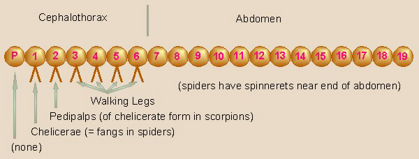
S-P. CHELICERATA
C. ARACHNIDA
spiders, scorpions |
0 |
4 prs, att. to cephalothorax, (chelicerae & pedipalps are m.p.) |
cephalothorax & abdomen |
 |
S-P. CRUSTACEA
C. MALACOSTRACA
crayfish, crabs, pillbugs |
2 pair |
5 prs incl. cheliped att. to cephalothorax, and swimmerets, m.p.,
incl. mandibles |
cephalothorax & abdomen |
|
S-P. ATELOCERATA
C. DIPLOPODA
millipedes |
1 pair |
many, 2 pr per apparent segment bec of fused segm., m.p. incl.
mandibles |
head and trunk segments, every two segments fused into one
apparent segment |
|
S-P. ATELOCERATA
C. CHILOPODA
centipedes |
1 pair |
many, 1 pr per segment, m.p. incl. mandibles & poison claw on next
segm. |
head and trunk segments |
 |
S-P. ATELOCERATA
C. INSECTA
insects |
1 pair |
3 pr, 1 pr. per thoracic segment, m.p. incl mandibles, some
abdominal |
head, three-segmented thorax, segmented abdomen
(wings are not appendages) |
 |
Origins of Arthropod Appendages:
The theoretical origins of arthropod appendages from an
earthworm-like ancestor are thought to include the following stages:
Earthworm-like Ancestor
↓
Clamworm-like Ancestor
↓
Peripatus-like Ancestor
↓
Various Arthropods
Generic Arachnid:
Generic Crustacean:
Generic Insect:
Note that, for example, the chelipeds (pincers) of crayfish
and the chelicerae (pincers) of scorpions are not the same. The chelipeds
of crayfish are appendages of segment #8, while the chelicerae of scorpions
are appendages of segment #1. Note that in both the arachnids and the insects,
segment #8 has no appendages at all. In crustaceans, segment #1 bears the
antennae, and in insects, has no associated appendages.
Classification of Crayfish:
For many years, crayfish were classified as:
Kingdom Animalia
Phylum Arthropoda
Subphylum Mandibulata
Class Crustacea
Subclass Malacostraca
Order Decopoda
Genus Cambarus
However, even though crustaceans have mandibles, many
biologists now feel
that this is due to convergent evolution rather than a close relationship to
other mandibulates and question whether crustaceans should be included
in Subphylum Mandibulata. In Crustaceans, the biting surface of the mandibles
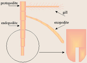
is thought to have evolved from an inner (mesal) bulge, or endite lobe
of the protopodite (note that the gills come from a similar outer
(lateral) bulge, the exite lobe) similar to the origins of the
mouthparts in the Trilobita and the Chelicerata). In all other mandibulates
(insects, centipedes, millipedes) the biting surface is modified from the
tips (the distal portion) of the endopodite.
Unlike the other mandibulates and similar to the Chelicerata,
crustaceans have a cephalothorax (cephalo = head), although
the head and thorax are often somewhat delineated. They have a telson
like trilobites had. Unlike any other arthropods, they have two pairs of
antennae: their first pair, the antennules, are homologous with the
antennae on insects and other mandibulates, while their second pair, the
antennae, have no homologous structures in insects and other
mandibulates. As mentioned above, crustacean
mandibles are slightly different in construction/origin from those of other
mandibulates, and they have more accessory mouthparts than any other
class.
Thus, an alternative classification scheme for the arthropods
is gaining acceptance, and more and more taxonomists are recognizing
Crustacea as a totally separate subphylum, unrelated to the rest of the
mandibulates.
Phylum Arthropoda
Subphylum Trilobita trilobites
Subphylum Chelicerata
Class Arachnida spiders, scorpions, etc.
Subphylum Crustacea crustaceans
Class Malacostraca
Order Decapoda
Genus Cambarus
Subphylum Atelocerata
Class Diplopoda millipedes
Class Chilopoda centipedes
Class Hexapoda insects
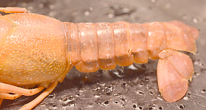
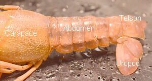
External Anatomy:
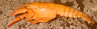
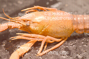
 Note the body regions: cephalothorax, covered by a
shell-like carapace (carapac = a covering, shield) on top,
and abdomen. Can you tell where the head and thorax join? Note the
abdomen, and see if you can find all seven abdominal segments.
The elongated, anterior portion
of the crayfishs cephalothorax is called the rostrum.
Note the body regions: cephalothorax, covered by a
shell-like carapace (carapac = a covering, shield) on top,
and abdomen. Can you tell where the head and thorax join? Note the
abdomen, and see if you can find all seven abdominal segments.
The elongated, anterior portion
of the crayfishs cephalothorax is called the rostrum.
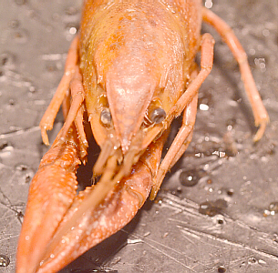
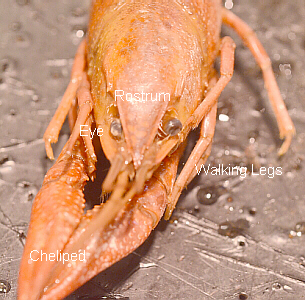
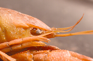
On the head, note the stalked, compound eyes. These
fit into grooves in the front of the head, and are often recessed into
them.

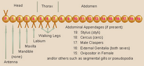
In many decapods (deca = ten, poda = foot:
referring to the walking legs), each segment of the abdomen bears a pair of
small appendages called swimmerets with the exception of the last
segment, called the telson. In crayfish, the penultimate segment bears a
pair of two-parted appendages called uropods (uro = tail).
The order gets its name from the ten walking legs (five pairs) located on the
thorax, including the cheliped (the pincer; cheli =claw,
ped =foot).
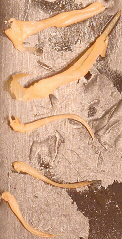
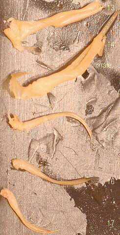
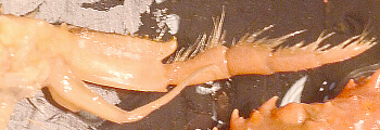
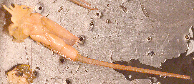
Observe all the appendages by carefully removing those from
the left side one at a time and arranging in order on the desk top
or a piece of paper for further study. It is easiest to do this starting
from the rear.
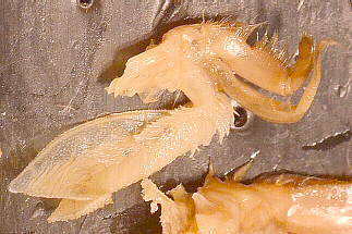
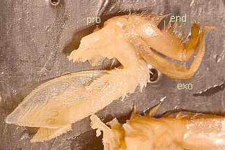
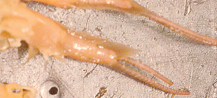
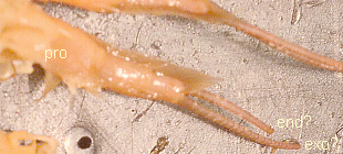
- The
telson is the last segment (#19) and does not have any appendages.

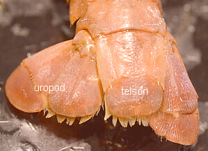
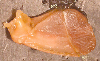
- The second-last
(penultimate) segment (#18) of many decapods has a broad, flat, two-part structure,
the uropod (uro = tail, poda = foot).
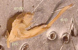
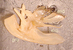
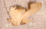
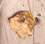
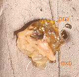
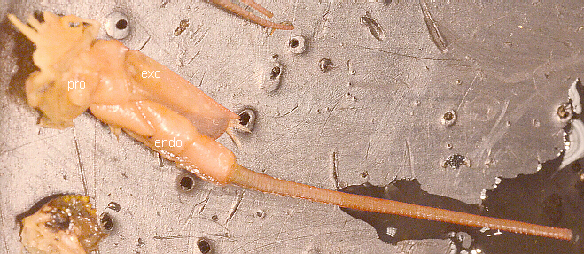
Note, in this and
other labeled drawings, below, exo refers to the exopodite, end refers
to the endopodite, and pro refers to the protopodite.
- The other
abdominal segments each bear a pair of small appendages called swimmerets.
Can you find all five pairs? Are they all identical or are some different?
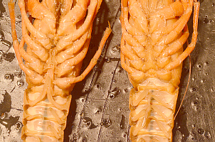
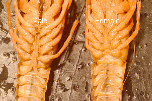
In crayfish, the swimmerets of the first two abdominal segments (13 & 14)
are larger in the male and modified for sperm transfer. Because of their use
in sperm transfer, some texts also refer to these specialized swimmerets as
gonopods. In females, the first two pairs of swimmerets are smaller
and look more similar to the remaining three pairs.
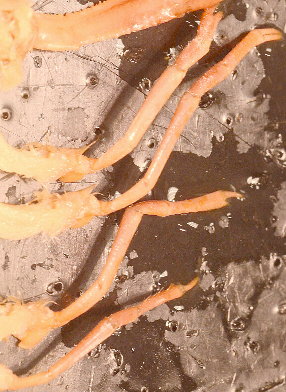
- The
thoracic region has five pairs of walking legs (deca = ten for the
ten legs of decapods), the front-most pair being modified as a cheliped
(cheli = claw, hoof; ped = foot) or pincer.
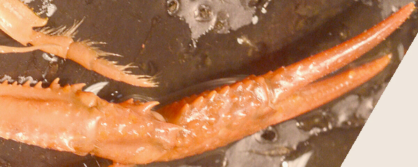
In some decapods,
each of the other four walking legs has a small pincer at the tip, while in
others, the walking legs have just one tip segment. Do the crayfishs walking
legs have one- or two-tipped ends? These legs are the appendages of segments
8-12.

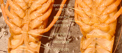
The openings of the females oviducts (ovi = egg) are
located at the bases of the third pair of walking legs (segment #10) with a
seminal receptacle (semin = seed, sperm, semen) between the
bases of the fourth pair of legs (segment #11). On the males, the openings
of the vasa deferentia (vasa = vessel, duct; deferens =
carry away) are located by the bases of the fifth pair of walking legs
(segment #12). As you remove these legs, note which have gills attached.
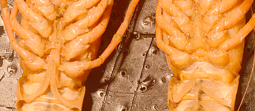
- The next
appendages are associated with the mouth. They are quite small,
flattened, and closely packed. The easiest way to remove each one is first
to determine exactly which is which, then grasp firmly at the base with a
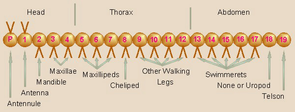
forceps and twist and pull it loose. From posterior to anterior, these are
the maxillipeds (maxill = the jaw, jawbone) (third, second,
first) on segments 7, 6, and 5 at the front of the thorax.

Third Maxilliped (Segment #7)
Second Maxilliped (Segment #6)

First Maxilliped (Segment #5)
Then, on segments
4 and 3 of the head, are the second and first maxillae, and on segment
2, the mandibles. The mandibles are heavy, grinding structures
associated with the mouth. Note if any of these appendages have
gills attached. Note in which direction the mandibles move.

Second Maxilla (Segment #4)
First Maxilla (Segment #3)
Mandible (Segment #2)
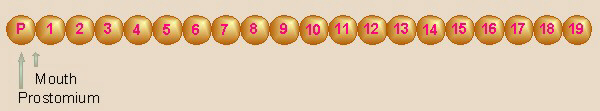
- The
appendages of segment 1 and the prostomium (pro = before, in
front of; stoma = mouth) are, respectively, the antennae and
the antennules (antenna = sailyard).
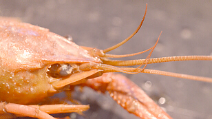
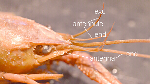
Antenna (Segment #1)
Antennules (Prostomium)
The external openings of
the green glands are located on the bases of the antennae (green gland
will be studied inside) although may be difficult to see.
Carefully cut away part of the carapace on the left side to
expose the gill chamber. Note the gills and the inner wall of
the gill chamber (the wall of the thorax). Note how some of the gills are
attached to the wall of the thorax and others to the bases of the appendages.
In the crayfish, the gills resemble feathers in shape. Count the number of
gills.
Internal Anatomy:
Beginning at the back edge of the carapace, cut two lines
forward, about ¼ to ½ inch (about 1 cm, or so) to each side of the middle.
Note: cut very shallowly because there are body organs located right below
where you will be cutting.
Gently lift off the flap, trying to remove only the chitinous exoskeleton.
The underlying epidermis may adhere to the exoskeleton or it may
remain covering the internal organs. If it remains, you will need to gently
remove it also. You may need to widen the opening further to view all the
organs. Also, continue these slits into the abdomen as far as feasible and
carefully remove the flap of exoskeleton.
 In the thorax, the first thing you should see dorsally is the
heart. You may be able to see small openings, ostia (os
= mouth), through which the blood enters the heart. At several points, you
may be able to see arteries leaving the heart. The circulatory
system is an open one, thus once the blood leaves the arteries, it
circulates throughout the body cavity. The membranous chamber in which the
heart lies is the pericardial sinus (peri = around;
cardio = heart).
In the thorax, the first thing you should see dorsally is the
heart. You may be able to see small openings, ostia (os
= mouth), through which the blood enters the heart. At several points, you
may be able to see arteries leaving the heart. The circulatory
system is an open one, thus once the blood leaves the arteries, it
circulates throughout the body cavity. The membranous chamber in which the
heart lies is the pericardial sinus (peri = around;
cardio = heart).
If your crayfish is a female, much of the thorax will probably
be filled with eggs (brownish). Below the heart, behind the
stomach are the reproductive organs. As mentioned, if the
ovaries (ova, ovo = egg) of the female are filled with eggs,
they will be easily recognizable, although the ovary walls will be invisible.
Those of a female without eggs as well as the testes of the
male may be difficult to distinguish from the digestive glands and may
even be partially imbedded in the glands. Generally, the reproductive organs
(the ovaries at least) are a darker, orange-brown color and tougher than the
digestive glands. The females oviducts extend toward
the bases of the third pair of walking legs while the males vasa
deferentia open between the fifth walking legs. Check a
crab/crayfish of the opposite sex to see the differences.
Intact, Unopened Stomach/Gizzard
Opened Gizzard with One
Grinding Structure Removed
Forward of (and perhaps slightly below) the heart is the
stomach. To see this, you may gently need to remove some of the eggs
and the heart. You may be able to insert a blunt probe through the
mouth and short esophagus (eso = within, inward;
phago = eat) into the stomach. In the crayfish, the stomach is
divided into anterior and posterior sections (crop and gizzard) by a
constriction bearing
grinding structures to shred the food as it passes. From the stomach, the
intestine continues to the end of the abdomen, but may be hidden in
the thorax by other organs. A pair of large, soft, yellowish structures
around the stomach (under the eggs) are the digestive glands. You may
remove the heart (and eggs), if not already removed, to see the digestive
glands better. They are connected to the intestine and aid in digestion and
absorption of food.
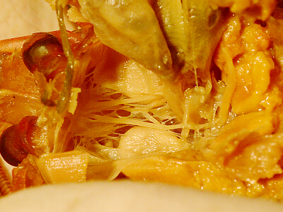
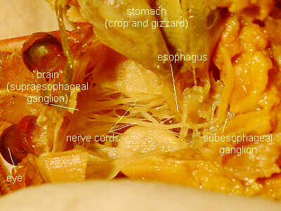
Side View of Nerve Cord Going Around Esophagus
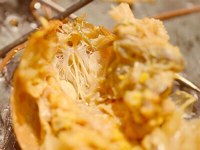
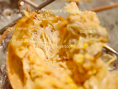
Rear View of Brain and Nerve Cord Going Around Esophagus
The nerve cord is located ventrally. It is a pair of
thin, whitish, fairly tough strings that can best be located by first
looking for the two halves that go around the esophagus. To follow the
nerve cord posteriorly, you may have to gently and carefully remove
abdominal muscles and/or push aside or remove the digestive
organs in the thorax. Note that there is a nerve ganglion
in each segment and that they are connected by the double-stranded nerve cord,
resulting in the ladder-like appearance of the nerve cord.
In the thorax, there are chitinous structures over the cord which will need
to be cut away to see it. Each of the posterior thoracic
segments bears its own ganglion, but those of the anterior thoracic and
posterior head segments (segments 3 through 7) are fused
to form one subesophageal (under the esophagus; sub = under,
beneath) ganglion (gangli = a knot on a string, swelling). The
two halves of the nerve cord continue anteriorly around each side of the
esophagus to the supraesophageal (over the esophagus; supra
= above, over, beyond) ganglion or brain, from which nerves
radiate to the eyes, antennae, etc.
Examine the various thoracic and abdominal nerve ganglia to
determine whether the those ganglia are the same or different in size. How
do you think the size of a ganglion correlates to the functions being
performed in/by that segment of the body?
In the anterior part of the body cavity, between the esophagus,
supraesophageal ganglion, and attachment of the antennae, are
the green glands. These excretory organs function similarly to our
kidneys in that they remove waste from the blood and discharge
it to the outside.
Copyright © 2010 by J. Stein Carter. All rights reserved.
Based on printed protocol Copyright © 1989 J. L. Stein Carter.
This page has been accessed  times since 18 Dec 2010.