Fungi
Background Information on Fungi
Fungi are eukaryotes and are heterotrophs that absorb (not
ingest) their food. They secrete digestive chemicals into the environment,
where the food is digested, after which they absorb the nutrients. Most
fungi are multicellular (yeast are secondarily unicellular). Fungi can be
saprophytes, parasites, or mutualistic symbionts.
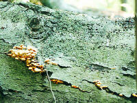
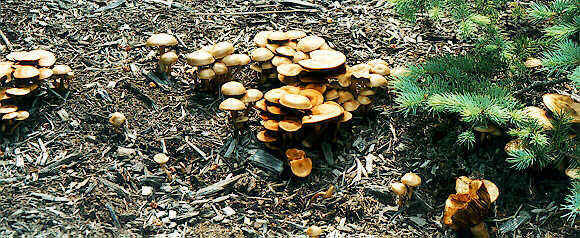
Saprophytes in Beech Log
Saprophytes
absorb nutrients from dead organic matter (dung, corpses, etc.). These are
important, necessary decomposers.
Parasites
absorb nutrients from the body fluids of a host organism, to the detriment
of the host.
Mutualistic
symbionts
absorb nutrients from a host, but reciprocate with some beneficial
function(s). For example, mycorrhiza (-ae) (rhizo = root) are
special fungi that live in/on the roots of plants, especially trees.
This mutualistic association of plant roots and fungi is beneficial to both
organisms because, through their digestive enzymes, the fungi help make
minerals available to the plant and help in water absorption (they are
smaller diameter than any of the tree roots) in return for organic food
from the plant. About 90% of all trees depend on micorrhizae and bare-root
trees often dont do as well because all of the smaller roots where the
micorrhizal fungi would normally live are pruned off. I have seen
suggestions in various gardening books that when bare-root trees are planted,
a couple shovelfuls of forest soil should be put around their roots to
inoculate them with the necessary fungi.
The body of a fungus is called a mycelium (pl =
mycelia) (myce = fungus; -ium = small). A mycelium is a tangled network of
filaments, each of which is called a hypha (pl. = hyphae; hypha
= web, weaving). Some species of fungi have their hyphae divided into
individual cells while others have hyphae that are multinucleate tubes
without individual cells. Growth of a mycelium (the hyphae therein) can be
very rapid, hence mushrooms (which are composed of densly-packed hyphae)
can pop up in a lawn overnight. Even solid-looking muchrooms are made of
masses of densely-packed hyphae.
The normal condition for fungal nuclei is 1n, and mitosis
happens slightly differently than in other groups of eukaryotes. Normal
fungal reproduction is asexual by just making spores that are disseminated
by wind or water. However, especially under adverse conditions, many fungi
also have some form of sexual reproduction with the formation of a different
kind of spores, and fungi are grouped/classified based on what type of
sexual reproduction they use.
A number of fungi are commercially important: yeast and all
the various edible mushrooms are fungi; blue, Roquefort, Camembert, and Brie
cheeses are made using Penicillium roqueforti or Penicillium
camemberti; while P. notatum is the source of penicillin. A
variety of other molds are used for drugs, cheeses, etc.; other edible
mushrooms are used in Oriental cooking; some mushrooms like truffles,
puffballs, morels are considered to be delicacies: and various strains of
the yeast, Saccharomyces cerevisiae, are used in making beer, wine,
andxfs bread.
On the other hand, a number of fungi are
pathogenic
and adversely affect humans. Some mushrooms are toxic so you should never
eat wild mushrooms unless you are absolutely, positively sure what they are.
Ergot of rye grows in rye and if infected rye is milled into flour
and ingested, people who eat that flour will ingest the chemical
ergotine, which is both toxic and hallucinogenic (and also contains
lysergic acid from which LSD is made). Ergotine can cause spasms and a
burning sensation. Currently, ergotine is used in very small (dilute)
amounts to stop postpartum bleeding.
Candida albicans is normally a a single-celled,
yeast-type fungus
which lives in our large intestines. Normally, it is a small part of
the intestinal flora and is kept in check by the good bacteria that live in
our colons. If for some reason it becomes necessary for a person to take
antibiotics, as we previously discussed, these antibiotics will kill the
good bacteria, allowing Candida and other such invaders to multiply.
Also, if the person eats too much sugar or other simple carbohydrates, these
will serve as food for the Candida, helping it to multiply, grow,
and/or travel to other parts of the body, where it is called vaginal yeast
infection (both partners must be terated simultaneously or theyll just
pass it back and forth) or thrush (if its in someones mouth and/or
throat). There is also some evidence that Candida can turn into a
mycelium with hyphae invading body tissues. To help fight against a yeast
infection, a number of people have suggested eating yogurt daily, especially
if a person is on antibiotics, and I read, somewhere, that women who consume
a cup of yogurt a day have less problems with yeast infections. If a
person has a number of bacterial infections, and antibiotics are frequently
prescribed and/or if a person is prone to yeast infections, that person
should insist that his/her doctor prescribe a fungicide (nystatin,
which goes by brand names like Mycostatin, or Nilstat, Nystex, etc.) along
with any antibiotics that are prescribed. From what Ive read, it is
strongly suggested that such a person also avoid foods either naturally or
artificially high in sugar and simple carbohydrates (grapes, bananas, pop,
candy, cookies, etc. white flour, white rice) which just serve as food for
the Candida, making things worse. Rather, this person should replace
these with a high fiber, adequate protein diet. There is also evidence that
taking garlic on a daily basis helps garlic is a known fungicide. I have
heard that some people put yogurt on an infected site and that some women
use a yogurt douche, but it would probably be a good idea to discuss these
options with your physician first. It is important to keep the infected
area as dry as possible, and open to fresh air if possible. If the site is
normally covered by clothing, wear cotton clothing because polyester holds
in body moisture women with vaginal yeast infections shouldnt wear nylons.
Fungal Classification
Like botanists, mycologists (myco = fungus;
-ology = to study) use the term Division instead of Phylum and
there are five Divisions in Kingdom Fungi: Zygomycota, Ascomycota,
Basidiomycota, Deuteromycota, and Lichens. Most of the fungi with which we
are familiar are Basidiomycetes or Ascomycetes. Almost all fungi reproduce
asexually by producing some kind of spores. The various Divisions are
separated by the type of sexual reproduction done by their members.
Examine, draw large,
labeled pictures of, and take notes on
each of the following examples of fungi. Optionally, observe wild fungi
and bring back samples for microscopic examination. Certain other wild fungi
like morels or blackleg can be brought back and sautéed. Also, examine
any plastic mounts and/or other live or preserved specimens that are
available.
-
Division Zygomycota:
-
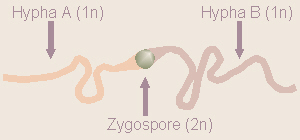
Diagram of Rhizopus Reproduction
(zygo = yoke; myco = fungus). In this group of fungi, sexual
reproduction produces a zygosporangium (containing zygospores) which
can remain dormant through unfavorable weather and release the spores when
weather is suitable. An example is Rhizopus stolonifera (rhizo
= root; pus = foot; stoloni = twig, shoot; fer = to bear,
carry), commonly known as black bread mold.
Hyphae are typically 1n. When opposite mating strains come into contact,
portions of the hyphae of each will grow toward each other, eventually
joining together to form a 2n zygote. The zygote develops a tough
protective coat and is then called a zygospore. Eventually, this
undergoes meiosis to form four 1n daughter cells.
-
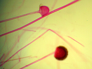
Prepared Slide of Rhizopus
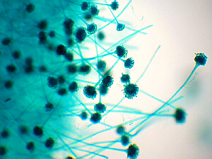
Examine the slide containing Penicillium, Aspergillus, and
Rhizopus (Carolina #B223). The Rhizopus is the one thats
stained red and bears large, knobby structures which contain spores.
-
Division Ascomycota (Ascomycetes):
-
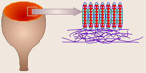
Diagram of Peziza Reproduction
(ascus = little sac, bag, bladder, hence commonly called sac fungi)
Fungi in this Division primarily
reproduce asexually by forming chains of spores called conidia
(conid = dust). In their sexual reproduction, a cup-like fruiting
structure is formed called an ascocarp (carpo = fruit). This
contains a number of asci (ascus), each of which contains eight
ascospores. The ascospores are always lined up in the order in which
they did meiosis, thus are used by some biologists to study meiosis.
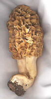
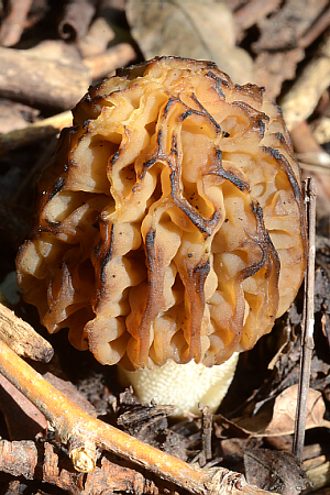
Examples of fungi in this Division include Peziza and other cup fungi,
as well as Morels
(Morchella) considered a delicacy and one of the few thats safe
to collect because few other things look like them. Yeast and Ergot are
also Ascomycetes. For many years, mycologists (people who study
fungi, myco = fungus) suspected that Penicillium belonged here,
and recently the genus was officially moved from the Deuteromycota to here.
When two opposite mating strains come into contact, a special structure, the
ascocarp (carpo = a fruit) is produced for sexual reproduction.
This consists of several specialized hyphae which terminate in structures
called asci (the sacs). Within each ascus, a zygote undergoes
meiosis to form four cells, each of which divides once more by mitosis. Thus
there is a total of eight ascospores in an ascus. Interestingly,
these have been used for genetic studies because they are lined up in the
order in which the chromosomes separated in meiosis.
-
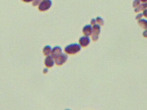
Prepared Slide of Aspergillus
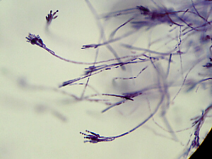
Prepared Slide of Penicillium
Examine the Aspergillus (stained blue) and the Penicillium
(stained purple) on the same slide mentioned above.
-

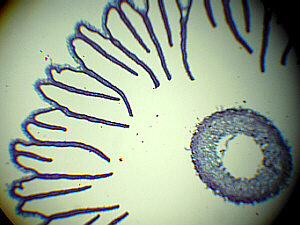
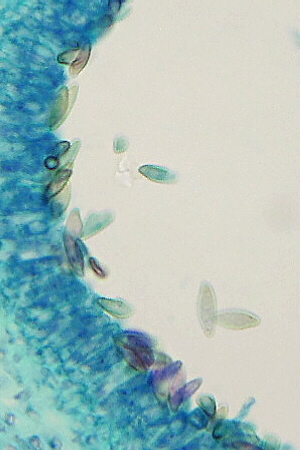
Peziza cross section

Peziza 8 Ascospores
 If available, cut a thin cross-sectional slice from a cup fungus
(Peziza) or morel and examine under the microscope to see the asci
and ascospores. If neither of those is available (as is likely in mid-winter)
examine the prepared slide of Peziza (Carolina #B255 pictured here),
instead. Notice that there are eight (8) ascospores per ascus.
If morels are available, sautée; and enjoy. If blue cheese or Camembert
is available, you may wish to taste some. Optionally, if there are any
left-over agar plates sitting around, P. roqueforti could be
cultured for later examination.
If available, cut a thin cross-sectional slice from a cup fungus
(Peziza) or morel and examine under the microscope to see the asci
and ascospores. If neither of those is available (as is likely in mid-winter)
examine the prepared slide of Peziza (Carolina #B255 pictured here),
instead. Notice that there are eight (8) ascospores per ascus.
If morels are available, sautée; and enjoy. If blue cheese or Camembert
is available, you may wish to taste some. Optionally, if there are any
left-over agar plates sitting around, P. roqueforti could be
cultured for later examination.
Follow this link (picture to the right) to view more photographs of
Ascomycota.
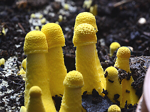
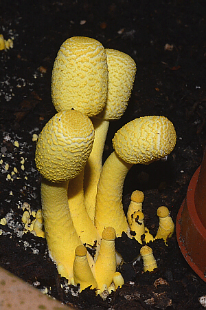
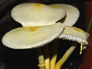
- Examples of other Ascomycota include:
Half-free Morel
Prepared Slide of Yeast Budding
-
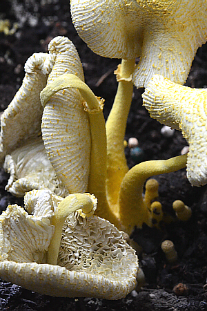
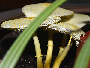
Division Basidiomycota (Basidiomycetes):
-
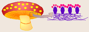
Diagram of Mushroom Reproduction
(basidium = little pedestal)
This Division includs things like mushrooms, shelf fungus, and puffballs:
You may have already observed and tasted some of these fungi in a previous
Biology lab class. A mushroom is actually a specialized reproductive
structure arising from a large underground mycelium. Technically, a
mushroom is a basidiocarp. The gills of mushrooms are lined with
special structures called basidia. Within each basidium a zygote
undergoes meiosis to form four nuclei. The basidium then grows four
appendages and a nucleus goes into each to make four basidiospores.
Often the mycelium of mushrooms starts
in one place (wherever the initial spore landed) and spreads out from there
in a circle. Around the edge of the circle, specialized, densely-packed
hyphae form fruiting structures each of which is called a basidiocarp
(or mushroom).
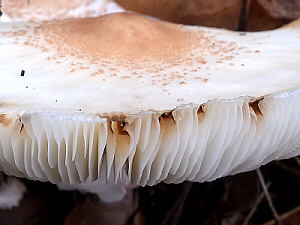
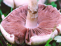
Mushroom Gills
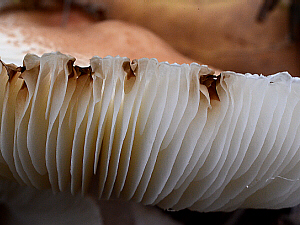
Gills Close-up
The gills of mushrooms are lined with basidia (sing.
= basidium) each bearing four basidiospores which result from meiosis.
Some members of this group (like Polyporus squamosus poly =
many; porus referring to the pores on the underside, squamo =
scale referring to the shaggy top side) have pores rather than gills. In a
puffball, spores are produced inside the puffball, then wait for a hole or
tear in it so they can get out. A ring of mushrooms forms at the outer edge
of the mycelium. Mushrooms can rapidly appear and use nutrients in the
area of the immediate growth ring, so the grass appears stunted and is called
a fairy ring (people thought the fairies were dancing and trampling
down the grass there).
-
-
Growth of Mushrooms:
| 1. Small Buttons | 2. Larger Buttons |
|---|
 |
 |
| 3. Caps Open Leaving Vellum Behind |
|---|
 |
| 4. Velum Left by Opening caps | 6. Decomposing Basidiocarps |
|---|
 |
 |
| 5. Caps, Stalk, and Velum |
|---|
 |
-
Coprinus Slice Through Cap

Coprinus Gills Close-up

Coprinus Basidiospores on Basidia
If available, cut a thin cross-sectional slice from mushroom gills
and examine under the microscope to see the basidia and basidiospores.
If no live specimens are available (as is likely in mid-winter) examine the
prepared slide of Coprinus (Carolina #B270 - pictured here), instead.
-
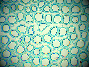
Polyporus Section
Polyporus Close-up of Basidiospores

Polyporus Section of One Pore
Polyporus is another member of the Basidiomycota. However, these do
not form gills like other mushrooms, but rather as their genus name might
suggest, make many, small, rounded pores in which the basidia and
basidiospores develop. Examine the prepared slide of a cross section
through a Polyporus (Carolina #B276).
 Draw and label the general structure of a mushroom, including cap, gills,
stalk, and annulus (= velum). If appropriate species of mushrooms are
available, sautée; and enjoy.
Draw and label the general structure of a mushroom, including cap, gills,
stalk, and annulus (= velum). If appropriate species of mushrooms are
available, sautée; and enjoy.
Follow this link (picture to the right) to view more photographs of
Basidiomycota.
- Examples of other Basidiomycota include:
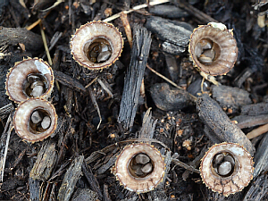
Birds-Nest Fungus
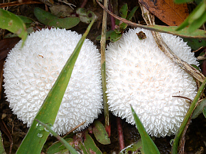
Small Puffballs
-
Division Deuteromycota
-
(deutero = second, i.e. other) This is a general, catch-all category
for fungi in which humans have not yet observed sexual reproduction (which
would qualify a fungal species to be classified in any of the other Divisions).
This Division is also called Fungi Imperfecti or the imperfect fungi
(botanists use perfect to refer to a plant with flowers with all their
sexual parts and functions while fungi do not have flowers, the method(s)
of sexual reproduction used by members of this Division is/are unclear to
human observers). Until
recently, Penicillium used to be classified as an imperfect fungus
because, while humans had observed asexual reproduction by means of
asexually-produced spores, no one had ever seen evidence of sexual
reproduction (fertilization and meiosis) in any species of Penicillium.
For many years, it was suspected that Penicillium belonged in the
Ascomycota, and recently it was officially moved to that Division.
-
This group is mentioned just because it fits in, here, but we will not be
examining them.
-
Lichens:
-
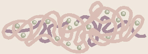
Diagram of Lichen Composition
The body of each species of lichen is made of a very specific mixture of
one kind of fungus (often an Ascomycete) and one kind of either a green alga
(Division Chlorophyta) or a bluegreen alga (Cyanobacteria). The fungus and
alga live in a symbiotic relationship,
that is they are closely associated with each other. More specifically,
their relationship is called mutualistic, because both benefit from
the relationship. The fungus holds water to keep the alga moist, digests
rock and makes the minerals available for both to use, and generally serves
as protection for the alga. The alga produces organic food for both by
the process of photosynthesis. The scientific names for lichens usually are
based on the type of fungus (each species of lichen consists of its own
kind of fungus and its own kind of alga). The scientific names for lichens
apply to each type of lichen there are not separate names for the fungus
and alga in a lichen. Lichens are often a grayish-green color, but may be
brightly colored. They are an important food source for a number of animals
(notably reindeer), while some species are used by humans for dye. Lichens
are good at colonizing bare rock and starting to break it down into soil.
Because lichens depend on rainwater as a source of moisture and air as a
source of CO2 from which to make sugar, they are extremely
sensitive to pollution and are among the first to die. Thus, these fungi
are indicator species for poor air quality.
-

Microscopic View of Lichen
Examine the prepared slide of a cross section of a lichen (Carolina #B294).
In general, as shown here, the fungus is stained blue, and the alga is
stained red.
Follow this link (picture to the right) to view more photographs of lichens.
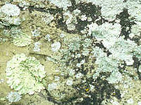
- Examples of other lichens include:
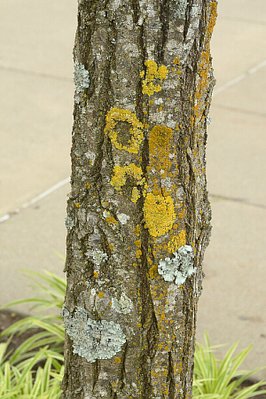
Lichens on Tree Trunk
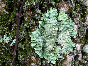
Lichen on Tree Trunk
Other Things to Include in Your Notebook
Make sure you have all of the following in your lab notebook:
- all handout pages (in separate protocol book)
- all notes you take as you read through the Web page and/or
during the introductory mini-lecture
- all notes and data you gather as you perform the lab
- labeled drawings (yours!) of all fungi examined
with all body parts labeled
- Zygomycota (Rhizopus)
- Ascomycota (Peziza)
- Ascomycota (Penicillium)
- Ascomycota (Aspergillus)
- Basidiomycota (Coprinus)
- Basidiomycota (Polyporus)
- Lichen
- any others that are available
- answers to all discussion questions, a summary/conclusion in your
own words, and any suggestions you may have
- any returned, graded pop quiz
Copyright © 2010 by J. Stein Carter. All rights reserved.
Based on printed protocol Copyright © 1995 J. L. Stein Carter.
Chickadee photograph Copyright © by David B. Fankhauser
This page has been accessed  times since 18 Dec 2010.