Protista
Background on Protista:
Some members of Kingdom
Protista
are unicellular, others are colonial, and yet others are multicellular.
Note that in the colonial forms, all the cells are similar with similar,
generalized functions, whereas in the truly multicellular species, the
body of the organism consists of a variety of types of cells, each type
with its own specialized function. Protists are the most primitive
eukaryotes (eu = good, well, true; karyon = nut, kernel,
nucleus); they have a true nucleus. They all need some kind of a
water-based environment, and may be found essentially anywhere there is
water, including both fresh and marine water, snow, damp soil and leaf
litter, and even inside polar bear hairs. All are aerobic (aer = air,
atmosphere, oxygen) and have mitochondria to do cellular respiration. Some
have chloroplasts and carry on photosynthesis while others are heterotrophs
(hetero = other, different; troph = food, nourish,
nourishment) and still others do both. Many protists have cilia
(cili = eyelash, small hair) and/or flagella (flagellum
= whip). Most of them reproduce or grow by mitosis, while some, but not all
of them, are capable of sexual reproduction and meiosis. Many can survive
periods of unfavorable conditions by forming cysts. Protists are a
major component of
plankton.
Several of the larger algae, especially various brown and red algae are
eaten by humans. Being marine, these algae are good sources of various
minerals, including iodine. Interestingly, the omega-3 fatty acids for
which fish oil is famous are not made by the fish, themselves, but by the
algae in their diets.
Protists are grouped into three major, unofficial categories
based on means by which they obtain nutrition. These are the Protozoa,
the Algae, and the Fungus-like Protists. For some reason,
botanists use the word Division to mean the same taxonomic level as
Phylum, and since, way back everything was lumped in as either a plant or
an animal, taxonomists who study Kingdom Protista (and those who study
Kingdom Fungi) also still use the word Division to mean Phylum, so for
example, when Division Rhizopoda is listed below, that means the same
thing as saying Phylum Rhizopoda.
Examine slide(s) and/or plastic mounts of the following
protists as available. Draw what you see and label parts such as nucleus,
chloroplasts, vacuoles, cilia, flagellum, etc. when visible. Note whether
the organisms are unicellular (uni- = one), colonial, or
multicellular.
One new piece of equipment we will be using for this lab is a
depression slide, also called a well slide. This is a special kind
of microscope slide with a lens-like depression on the top surface. When
making wet mounts of larger microscopic organisms, this gives them more
space to swim and not get squashed while you observe them than would be
possible with a regular microscope slide.
Protozoa: (Animal-Like Protists)
These protists are animal-like, especially in their nutrition.
They ingest their food by
phagocytosis.
Some have mouth-like structures into which the prey is put while others use
pseudopodia
to move and to engulf prey. Typical prey include bacteria and other smaller
one-celled organisms.
- Division
Rhizopoda:
-
 (rhizo =root; poda = foot)
An example of a member of this Division is genus
Amoeba
(amoeb = change),
a fresh-water dweller. Protists in this group are unicellular and have
pseudopodia. Some secrete shells around themselves, while others do not.
None of them have flagella, cilia, or meiosis.
Entamoeba histolytica
is a
parasitic
form that causes
amoebic dysentery.
These colonize the colon and feed on bacteria, causing symptoms that range
from mild diarrhea to dysentery. Typically periods of watery diarrhea, often
containing blood, may alternate with constipation, and often there is
flatulence and abdominal cramping. Entamoeba can be directly spread
(anal sex), or indirectly spread (by drinking contaminated water). Fresh
fruits and vegetables may be unsafe if fertilized with human feces, watered
with contaminated water, or prepared by a person with it on his/her hands.
(rhizo =root; poda = foot)
An example of a member of this Division is genus
Amoeba
(amoeb = change),
a fresh-water dweller. Protists in this group are unicellular and have
pseudopodia. Some secrete shells around themselves, while others do not.
None of them have flagella, cilia, or meiosis.
Entamoeba histolytica
is a
parasitic
form that causes
amoebic dysentery.
These colonize the colon and feed on bacteria, causing symptoms that range
from mild diarrhea to dysentery. Typically periods of watery diarrhea, often
containing blood, may alternate with constipation, and often there is
flatulence and abdominal cramping. Entamoeba can be directly spread
(anal sex), or indirectly spread (by drinking contaminated water). Fresh
fruits and vegetables may be unsafe if fertilized with human feces, watered
with contaminated water, or prepared by a person with it on his/her hands.
-
Make a wet mount of live Amoeba in a depression slide (well
slide), put a coverslip on top, and then examine under the
microscope. Notice its pseudopodia (pseudo = false) and any internal
organelles that are visible. They are unicellular.
- Division
Ciliophora:
-
 (cili = eyelash, small hair; phora = to bear, carry)
An example of an organism in this Division is
Paramecium
(paramec = oblong, oval).
These protozoans are solitary, fresh water organisms and use cilia to move.
They have probably the most complex structure and organization of all cells.
Rather than one nucleus, they have a larger macronucleus and several
smaller micronuclei. They use a form of sexual reproduction called
conjugation in which some of the micronuclei are exchanged between
the two individuals involved.
(cili = eyelash, small hair; phora = to bear, carry)
An example of an organism in this Division is
Paramecium
(paramec = oblong, oval).
These protozoans are solitary, fresh water organisms and use cilia to move.
They have probably the most complex structure and organization of all cells.
Rather than one nucleus, they have a larger macronucleus and several
smaller micronuclei. They use a form of sexual reproduction called
conjugation in which some of the micronuclei are exchanged between
the two individuals involved.
-
Make a wet mount of live Paramecium in a depression slide, put a
coverslip on top, and then examine under the
microscope. Look for a Paramecium that is holding relatively still.
Notice its numerous cilia, the oral groove leading to its
mouth, the contractile vacuole (which serves a kidney-like function in
that it works to expel/excrete excess water from the Paramecium),
macronucleus, micronuclei, food vacuoles (macro = large, long;
micro = small). Note that while many of these structures will be
visible on a prepared slide, a number of them may not be visible or as
visible in living Paramecium.
They are unicellular.
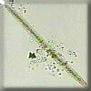
- Division
Zoomastigophora:
-
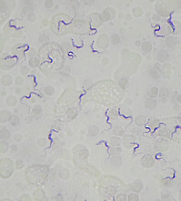
Trypanosoma gambiense
(zoo = animal; mastigo = whip)
This Division contains some organisms which are free-living, others which are
symbionts,
and yet others which are parasites. An example of a symbiotic member of this
Division is the protozoans which live in the gut of termites and digest
cellulose in the wood the termites eat. An example of a parasitic form would
be
Trypanosoma gambiense
(trypano = a hole, bore; soma = body; gam = marriage;
bios = life; -ense = of, belonging to),
which causes African sleeping sickness and is spread by the bite of
the tsetse fly. Symptoms include irregular fever, general swelling of the
lymph nodes, skin eruptions, and areas of painful local swelling. Eventually
CNS symptoms like tremors, headache, apathy, and convulsions appear and
become worse, leading to eventual coma and death. Early on, the parasites
are found in blood and lymph, but later only in the persons cerebrospinal
fluid.
-
Examine a prepared slide (Carolina # PS-310) of blood infected with
T. gambiense. Notice the round/oval RBCs.
The small, black, wiggly things are them. Notice the whip-like flagellum.
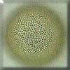
- Division
Apicomplexa:
- These are all parasites and form tiny, infectious spores. All have
complex life cycles. An example is
Plasmodium vivax,
which causes
malaria,
for which certain species of mosquitoes are the secondary host. It is also
possible to become infected with Plasmodium parasites from a
transfusion from an infected person or if a drug addict shares a syringe with
an infected person. One stage in this complicated life cycle grows in the
mosquito, the next stage in the newly-infected persons liver, and the next
stage invades the persons red blood cells, rupturing the RBCs as the
parasites leave to invade other cells. Symptoms include cyclical
alternating chills, fever, and sweating which at first, can be mistaken for
flu. While usually less than 1% of the RBCs are infected, often malaria
causes anemia due to the smaller number of RBCs. Often the spleen and liver
become enlarged as they try to deal with the dying RBCs. Malaria is treated
with extract from the quinine tree. Remember that people with sickle-cell
are more resistant because when a malaria parasite enters a RBC, the RBC
sickles, killing the parasite, thereby preventing it from multiplying and
spreading.
-
This group is mentioned just because it fits in, here, but we will not be
examining them.
Algae: (Plant-Like Protists)
These protists are photosynthetic; their nutrition is
plant-like. Almost all of them have chlorophyll A, most have chlorophyll C,
but only a few have chlorophyll B. They also have a variety of carotenoids
and other pigments, and frequently they are grouped into Divisions based on
similarities in pigments.
- Division
Euglenophyta:
 (eu = good, well, true; gleno = pit, socket; phyta = plant)
Probably the best-known example of this Division is genus
Euglena.
Each of these organisms has a
flagellum
on its
anterior
end, and this is used to propel the organism. They have chloroplasts and,
when in the light, do photosynthesis. If they are not in the light, they
can also obtain nutrition by phagocytosis. To help them sense light (which
they then move toward), Euglena have a light-sensitive eyespot or
stigma
near their anterior ends. This is not a true eye, in that it cannot do any
image formation, but rather it is a
photoreceptor
which senses the light level in the organisms environment.
(eu = good, well, true; gleno = pit, socket; phyta = plant)
Probably the best-known example of this Division is genus
Euglena.
Each of these organisms has a
flagellum
on its
anterior
end, and this is used to propel the organism. They have chloroplasts and,
when in the light, do photosynthesis. If they are not in the light, they
can also obtain nutrition by phagocytosis. To help them sense light (which
they then move toward), Euglena have a light-sensitive eyespot or
stigma
near their anterior ends. This is not a true eye, in that it cannot do any
image formation, but rather it is a
photoreceptor
which senses the light level in the organisms environment.
-
Make a wet mount of live Euglena in a depression slide, put a coverslip on
top, and then examine under the microscope.
Notice the flagellum at the anterior end, the reddish stigma
(stigma = spot) or eyespot (also at the anterior end), the nucleus,
and the green chloroplasts (chloro = green; plast = formed or
molded). These are autotrophs or heterotrophs (auto = self;
hetero = other, different; troph = food, nourish,
nourishment), depending on presence/absence of light. If you can find one
thats holding fairly still, you may be able to see the shadow of its
flagellum moving. Notice how, as they swim, they roll over, change shape,
and spin in circles. They are unicellular.
- Division
Chlorophyta (Green Algae):
- (chloro = green, phyta = plant)
These protists are also known as the green algae. Their chloroplasts
and the pigments therein are similar to plants (this is about the only group
of algae with chlorophyll B), thus it is thought that the green algae may be
the evolutionary ancestors of plants. Various species of green algae may be
found in a variety of environments including both fresh and salt water, damp
soil, the surface of snow, and within other organisms (lichens, hydra, polar
bear hair).
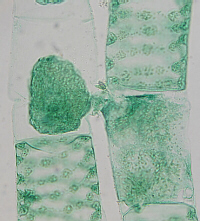
-
Ulva
-
(ulva = a sedge)
This is called Sea Lettuce. Ulva is truely multicellular, with
a division of labor among the various cells, and it is macroscopic. The
body is two cells thick, and there is a specially-modified holdfast
to anchor the organism to the ocean floor. Its life cycle includes both
1n and 2n stages (see below).
-
Observe a plastic mount of Ulva. Notice the large size compared
to other algae. Note life cycle as described in your lecture textbook.
The 2n sporophyte and 1n gametophyte generations look the same.
The body is two cells thick.
-
Volvox
-
 (volv = roll, turn)
These are colonial and often contain darker green daughter colonies
inside. Each cell posesses two flagella, enabling the colony to be
mobile. There is an intercellular matrix holding the colony of
cells together.
(volv = roll, turn)
These are colonial and often contain darker green daughter colonies
inside. Each cell posesses two flagella, enabling the colony to be
mobile. There is an intercellular matrix holding the colony of
cells together.
-
Make a wet mount of live Volvox in a depression slide, put a coverslip on
top, and then examine under the microscope.
These are colonial with a clear intercellular matrix holding the
cells together. All cells are similar, and there is no division of labor.
The colonies are mobile due to the presence of two flagella on each cell.
Notice the darker green daughter colonies inside. They also are capable
of a form of sexual reproduction in which eggs and sperm are produced.
-
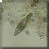
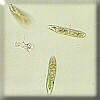
Closterium
-
 (closter = thread, yarn)
This is a member of the sub-group called the Desmids. Some
desmids form colonies, but Closterium is solitary. Its nucleus
is in the
center with a cone-shaped chloroplast on each side. Each chloroplast
contains a series of starch-storage organelles called
pyrenoids
pyren = a fruit stone; -oid = like, form).
In living Closterium, each end of the cell bears a small vacuole
containing several gypsum grains which dance by Brownian motion.
(closter = thread, yarn)
This is a member of the sub-group called the Desmids. Some
desmids form colonies, but Closterium is solitary. Its nucleus
is in the
center with a cone-shaped chloroplast on each side. Each chloroplast
contains a series of starch-storage organelles called
pyrenoids
pyren = a fruit stone; -oid = like, form).
In living Closterium, each end of the cell bears a small vacuole
containing several gypsum grains which dance by Brownian motion.
-
Make a wet mount of live Closterium in a depression slide, put a
coverslip on top, and then examine under the microscope. Look for the
nucleus in the center and the two, cone-shaped chloroplasts (overall, it
looks rather like two, green, ice-cream cones sharing one scoop of ice
cream between them). Note the row of circular pyrenoids within the
chloroplasts. Examine the vacuole on one of the tips of the cell under
high power to see the jiggling gypsum grains. These are unicellular.
-
Spirogyra
-
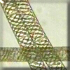 (spiro = breathe, spiral, coil; gyra = round, turning, a circle)
These are colonial, being organized into long filaments. Each cell
contains a spiral chloroplast with pyrenoids (used to store starch) and
a nucleus. They reproduce by
conjugation
a type of sexual reproduction in which the contents of the male gamete
cell go over into the female cell.
(spiro = breathe, spiral, coil; gyra = round, turning, a circle)
These are colonial, being organized into long filaments. Each cell
contains a spiral chloroplast with pyrenoids (used to store starch) and
a nucleus. They reproduce by
conjugation
a type of sexual reproduction in which the contents of the male gamete
cell go over into the female cell.
-
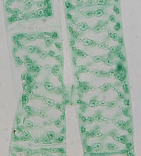
conjugation in Spirogyra
conjugation in Spirogyra
-
Make a wet mount of live Spirogyra in a depression slide, put a
coverslip on top, and then examine under the microscope. Notice the
filamentous colonies (which probably made it difficult to make a wet
mount). Notice the spiral (helical) chloroplasts which periodically
widen to accomodate the presence of pyrenoids. You may or may not be
able to find the nucleus in each cell. Notice the cell walls that
delineate the individual cells.
Examine a prepared slide (Carolina # B65) of conjugation in Spirogyra.
Conjugation (con = with, together; juga = a
yoke; -tion = process of, action of) is a type of
sexual reproduction in which the contents of the male gamete cell go
into and unite with the female cell.
-
Chlamydomonas
-
 These are unicellular and contain an eyespot (stigma), a chloroplast, two
flagella, and a nucleus.
These are unicellular and contain an eyespot (stigma), a chloroplast, two
flagella, and a nucleus.
-
In the past, we have examined live Chlamydomonas, but due to current time
constraints, we will not be looking at them this year. They are small,
fast-moving, green spheres with two flagella.
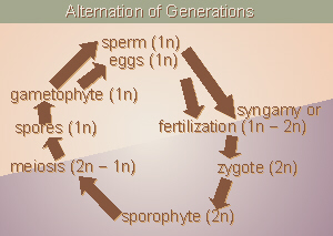 Many green algae, especially the multicellular ones, have both sexual and
asexual stages in their life cycles, thus we must introduce the idea of
Alternation of Generations we discussed along with meiosis. When we
first discussed Alternation of Generations, we looked at a very simple
diagram in which adults produced 1n gametes by meiosis, and those gametes
joined by syngamy to form a new 2n generation. In reality in algae and
plants, there are a few more stages in the process, thus we now need to
revisit this cycle. The 2n generation, which in humans is called an adult,
in algae and plants is called a
sporophyte
because it produces spores. Within specialized reproductive structures
in/on the bodies of the sporophyte, meiosis occurs to reduce the chromosome
number from 2n to 1n, thus the spores which are produced are 1n. What is
very significant, here, is that meiosis produces 1n spores, not
gametes. Each spore germinates and grows into a new, independent, 1n
organism (which often looks totally different than the 2n generation).
These 1n organisms are called
gametophytes
because they produce the gametes (eggs and sperm), which are still 1n. Then,
as weve seen in the past, an egg and sperm unite by
syngamy
(fertilization) increasing the chromosome number from 1n to 2n, and forming a
zygote
which is 2n. The zygote grows into the sporophyte, and the cycle starts over.
Various of the green algae go through this cycle as do members of the next
two groups, the brown and red algae. Plants also go through this same cycle
with some interesting modifications we will discuss later.
Many green algae, especially the multicellular ones, have both sexual and
asexual stages in their life cycles, thus we must introduce the idea of
Alternation of Generations we discussed along with meiosis. When we
first discussed Alternation of Generations, we looked at a very simple
diagram in which adults produced 1n gametes by meiosis, and those gametes
joined by syngamy to form a new 2n generation. In reality in algae and
plants, there are a few more stages in the process, thus we now need to
revisit this cycle. The 2n generation, which in humans is called an adult,
in algae and plants is called a
sporophyte
because it produces spores. Within specialized reproductive structures
in/on the bodies of the sporophyte, meiosis occurs to reduce the chromosome
number from 2n to 1n, thus the spores which are produced are 1n. What is
very significant, here, is that meiosis produces 1n spores, not
gametes. Each spore germinates and grows into a new, independent, 1n
organism (which often looks totally different than the 2n generation).
These 1n organisms are called
gametophytes
because they produce the gametes (eggs and sperm), which are still 1n. Then,
as weve seen in the past, an egg and sperm unite by
syngamy
(fertilization) increasing the chromosome number from 1n to 2n, and forming a
zygote
which is 2n. The zygote grows into the sporophyte, and the cycle starts over.
Various of the green algae go through this cycle as do members of the next
two groups, the brown and red algae. Plants also go through this same cycle
with some interesting modifications we will discuss later.
- Division
Dinoflagellata:
- These are abundant in plankton, occasionally occurring in large numbers.
They can occasionally become so numerous that the water looks red, thus this
algal bloom (meaning there are large numbers of them, having nothing
to do with flowers, which they do not have) is called Red Tide.
Because Dinoflagellates are toxic to humans, it is not safe to eat
shellfish (clams, etc.) collected where Red Tide is occurring (the
Protists get inside the clam shell and cannot be easily removed).
Dinoflagellates are
bioluminescent,
that is, they are able to produce light like lightening bugs, and at night
during Red Tide, the crests of the ocean waves appear to glow in the dark.
-
This group is mentioned just because it fits in, here, but we will not be
examining them.
- Division
Phaeophyta (Brown Algae):
- (phaeo = dusky, brown; phyta = plant)
These organisms are commonly known as the brown algae. They are
multicellular and live in marine, temperate zone, costal areas. They all
have a form of sexual reproduction with alternation of generations. One
member of this Division with which you may be familiar is Kelp, which
actually can be any of several species of seaweed in the genera Fucus
and/or Laminaria (fucus = seaweed; lamina = a thin
sheet, layer, plate). Brown algae are used in many cultures as human
food, and are good sources of iodine. We need iodine for our thyroid glands,
and if a person doesnt get enough iodine in his/her diet (most commonly in
inland areas where iodine is not added to salt), the thyroid gland enlarges
in an attempt to keep making enough thyroid hormone (which doesnt do any
good because what its lacking is the iodine needed to make the hormone).
This enlarged thyroid is called a goiter.
Laminaria
also has an interesting gynecological (gyneco = a woman,
female; logy = to study, the study of) use. If a woman is scheduled
for some medical procedure for which the doctor needs access to the inside
of her uterus, often a day or so beforehand, rolled-up, dried pieces of
Laminaria are inserted into the opening of the womans cervix. As
the seaweed absorbs water from her body fluids, it gently and slowly expands,
gradually stretching the cervix. Thus, by the time her surgery is scheduled,
her cervix has been dilated slowly and gently rather than the doctor having
to forcibly and quickly (thus painfully) stretch the cervix open minutes
beforehand. Iodine was first discovered by distillation
from Fucus. Carrageen from Irish Moss (Chondrus crispus) is
often used as a gelatin substitute and thickener for ice cream and salad
dressings. Agar-agar (or commonly, just agar), also used as a gelatin
substitute, is derived from Gelidium spp..
-
Examine (and taste?) any edible brown algae that are available.
Examine available plastic mounts. These have a multicellular body that is
differentiated/organized to serve a variety of functions.
- Division
Rhodophyta (Red Algae):
- (rhodo = a rose)
These are called the red algae. They also are multicellular and
marine-dwelling, but are more typically found in tropical zones and deeper in
the ocean. They also go through alternation of generations.
Many of these sea vegetables are used by people in other cultures and are
quite high in minerals such as iodine (which is low to absent in terrestrial
foods, hence our iodized salt). Dulse is one species which is commonly
consumed by humans. Nori is dried and (usually) toasted Porphyra
tenera, another seaweed.
-
Examine (and taste?) any edible red algae that are available.
Examine available plastic mounts.
Slime Molds (Fungus-Like Protists)
- Division
Myxomycota (Plasmodial Slime Molds):
- (myxo = slime, mucus; myco = fungus)
These organisms are called slime molds. They are fungus-like in their
nutrition in that they absorb nutrients from their environment. Their
body structure is unusual in that the nuclei undergo mitosis, but there is
no cytokinesis, so there are no individual cells with one nucleus each.
Rather, the body is a giant, multinucleate mass of cytoplasm. Slime molds
are mobile: they move by amoeboid movement, in other words, like a
giant Amoeba with giant pseudopodia. They live in decayed wood and move
around in between the fibers, ingesting bacteria, etc. by phagocytosis.
Slime molds are often brightly-colored (yellow or orange).
-
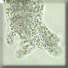
Physarum sp. is a multinucleate, macroscopic mass of cytoplasm, often
bright yellow in color. It
travels by ameboid movement. As mentioned, it has no cell walls, no
internal division into cells. It eats by phagocytosis (phago = to
eat; cyto = cell). See your lecture textbook for an explanation of
its life cycle.
Physarum can be cultured on a plain agar plate (just agar and water)
sprinked with oat flakes (oatmeal). The agar medium supplies water/moisture,
and bacteria that grow on the moist oat flakes serve as food for the
organism.
Examine a Physarum organism. Note that since this
is one, whole, macroscopic organism, you should NOT try to carve out
samples of it, but rather, just look at it. Your instructor may, optionally,
place it under a microscope so your class can view the cytoplasmic streaming.
Also, as time permits, examine pond water to try to identify
whatever protists you can find.
Draw large illustrations of and describe each of the organisms you examine
and label all identifiable parts. Take notes on any
significant/interesting structures, etc.
Other Things to Include in Your Notebook
Make sure you have all of the following in your lab notebook:
- all handout pages (in separate protocol book)
- all notes you take as you read through the Web page and/or
during the introductory mini-lecture
- all notes and data you gather as you perform the lab
- labeled drawings (yours!) of all protists examined
with all organelles/body parts labeled
- Amoeba
- Paramecium
- Trypanosoma
- Euglena
- Volvox
- Closterium
- Spirogyra
- Fucus
- any other brown/red algae
- slime mold (Physarum)
- answers to all discussion questions, a summary/conclusion in your
own words, and any suggestions you may have
- any returned, graded pop quiz
Copyright © 2010 by J. Stein Carter. All rights reserved.
Chickadee photograph Copyright © by David B. Fankhauser
This page has been accessed  times since 18 Dec 2010.