2-D says, I have a circulatory system, too, but mine is different than yours. You have all sizes of arteries, veins, and capillaries, but I dont have those. I have a heart that runs along the top, center of my abdomen and pumps my blood forward, and an aorta that comes out of the front end of my heart to carry the blood forward to my head, but thats about it. The other end of my aorta just opens into my body cavity near my head, and my blood flows over and around all of my body organs from there. Eventually, it finds its way back into my heart, and is pumped forward into my aorta, again. Another difference is that while you use your blood to carry oxygen to all your body organs, my tracheal system takes air straight to my organs, so my blood does not need to carry oxygen.
All animals must exchange materials with their environment, including nutrients and wastes, O2, CO2, etc., thus need a system that will do this. The more complex the organism, the more complex this system must be. Arthropods, like insects and spiders, have an open circulatory system, in which the blood is pumped forward by the heart, but then flows through the body cavity, directly bathing the internal organs. Vertebrates, like humans, have a closed circulatory system in which the blood stays in the circulatory system as it circulates, and chemicals are exchanged by diffusion. Our system is also called our cardiovascular system, and is composed of our heart plus our arteries and veins. In a persons heart, the atria (plural of atrium) receive blood from the veins and the ventricles send blood to the arteries. As the arteries become more finely divided, they are called arterioles. The finest divisions of our vascular system are called capillaries. As the vessels get larger again, the smallest are called venules which join and enlarge to form veins. Note that the distinction between arteries and veins is by direction of blood flow, not oxygen content. Veins carry blood toward the heart and Arteries carry it Away from the heart. Because of this, not all arteries carry oxygenated blood. The two major exceptions, in which arteries are carrying deoxygenated blood are the pulmonary artery which carries deoxygenated blood from the heart to the lungs (to pick up oxygen there) and the umbilical arteries which carry deoxygenated blood away from the babys body to the placenta (to pick up oxygen there). We have double circulation: we have a separate pulmonary circuit to the lungs and a systemic circuit to the body.
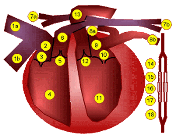
This illustration is orientated as thought you were looking at this heart in another person standing in front of you. The path of blood flow in a human, then, is as follows:
The atrioventricular and semilunar valves prevent backflow as the heart contracts. Defects in any of these that allow some blood to leak backwards cause distinctive sounds through a stethoscope, thus are called heart murmurs.
The sinoatrial node controls the heart beat. This natural pacemaker is located in the upper wall of the right atrium, and is composed of muscle tissue that sends electrical impulses to the rest of both atria to contract. The impulse then spreads to the ventricles, causing them to contract. The heart cycle involves three phases:
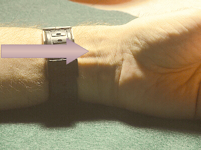
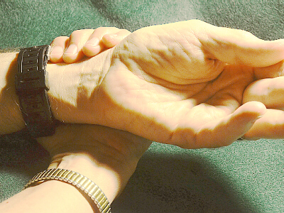
Determining Pulse
The rate of contraction is the heart rate. An
unborn babys heart starts beating when it is about four weeks old (the
mothers period is two weeks late, and shes just beginning to wonder if she
might be pregnant, but doesnt know for sure). A newborns heart rate is
around 135 to 140 beats per minute (bpm). By age 15 to 30, the rate
decreases to about 65 to 75 bpm, then speeds up slightly as the person ages.
The pulse is a wave of contraction of the artery walls (which roughly
corresponds to the heart rate) as blood is forced into the arteries. Pulse
is usually measured using the radial artery (the one along the radius).
To find your pulse, rest your right arm in the palm of your left hand. Curl
the fingers of your left hand up around the thumb side of your right wrist.
Place several fingers of your left hand along and just to the outside (thumb
side) of the tendon that runs along your wrist. With gentle pressure, you
should be able to feel your pulse.
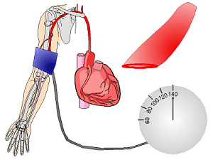
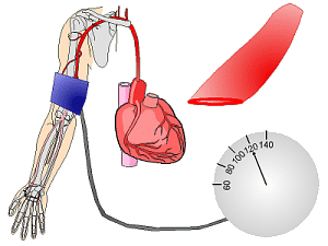
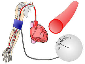
Determining Blood Pressure
(clipart edited from Corel Presentations 8)
Blood pressure is maximum during systole, when the heart is pushing,
and minimum during diastole, when the heart is relaxed. In a living person,
the blood pressure doesnt go to zero because the thick, elastic artery walls
exert pressure on the blood. A
sphygmomanometer
is the instrument used to determine BP. The artery used to determine BP is
the
brachial artery,
which runs down the upper arm, splitting into the radial and ulnar arteries
near the elbow. The cuff of the sphygmomanometer is wrapped around the arm
just above the elbow and pumped up to block off blood flow (the pressure
exerted by the cuff is higher than the systolic pressure). The pressure in
the cuff is gradually decreased, and when it equals the persons systolic
pressure, the heart can force blood under the cuff, and a sound is heard as
the pulses of blood surge under the cuff. As the pressure in the cuff is
lowered, when it equals the diastolic pressure, blood can flow freely, so
the sound disappears (not enough pressure is exerted by cuff to restrict
blood flow). Thus, by listening for the first sound, and when the sound
becomes faint, while watching the pressure indicator on the sphygomomanometer,
it is possible to determine someones blood pressure. Typically, when you
go to the doctors office, one of the first things that is done to you is
that someone (a nurse?) takes your blood pressure. I have frequently had
the experience that when I ask what the results were, I initially get the
answer Its OK. Heres a tip: you, not they, are in charge of
your health. The only way you can educate yourself to how your body works
is to keep re-asking the question until you get a real answer. You need to
know the actual numbers to be able to evaluate if things have changed or are
good or bad. Be persistent and eventually theyll tell you what your BP is.
A neonates BP is around 80/45 mm Hg meaning that the systolic pressure is equivalent to air pressure that will support a column of mercury 80 millimeters high in a barometer, and the diastolic is equivalent to the air pressure that will support a column of mercury 45 millimeters high. For adults in their 20s, 120/80 mm Hg is considered average for a male and 115/75 mm Hg for a female, thus the accepted average is said to be 120/80 mm Hg. With age, the arteries become less elastic (due in part to undesirable lipid deposits in their walls), so the BP rises. Hypertension is when the BP is too high. There are two ways this could happen: either the systolic pressure is greater than 145 to 160 mm Hg and/or the diastolic is greater than 90 to 100 mm Hg. Many researchers feel that major contributing factors appear to include the amounts of salt, cholesterol (and other lipids), and sugar in ones diet and the amount of exercise the person gets. Frequently, diuretics are prescribed to try to remove water from the persons blood, thus lowering the blood volume and hopefully thereby, the BP. However, many diuretics also remove potassium (and other beneficial minerals?) from the persons system, and if serum potassium levels are not carefully monitored and go to low, this could cause a heart attack!
A thrombus is a blood clot (platelets and fibrin) which forms within a vessel and blocks the blood flow. These can result from surgery or from conditions like atrial fibrillation. An embolus is a moving thrombus which may get stuck somewhere. If thrombi or emboli lodge in an artery supplying blood to the heart, this can cause a coronary embolism or heart attack or myocardial infarction. If one of these becomes lodged in an artery in the lungs, it is also a life-threatening pulmonary embolism, and if in the brain, a cerebral (or cerebellar) embolism or stroke or cerebrovascular accident (CVA).
A hemorrhage is bleeding, especially profuse, and can be severe if internal.
A hematoma is a local swelling or tumor filled with blood; a bruise, especially a large one. Sometimes, if the injury is extensive, it can calcify as it heals, leading to a hard lump (which may need to be surgically removed).
Hemorrhoids are dilated or varicose veins in the anal area. Typically, these are caused not enough fiber in diet causing the feces to be very hard so the person has to strain to pass them. Increasing the amount of fiber in ones diet can help prevent hemorrhoids and possibly aid in healing mild cases. Because vitamin C is necessary for collagen synthesis, it is necessary for strong capillary walls (one of the first signs of a vitamin C deficiency is easy bruising), so that along with the bioflavonoid rutin (found in buckwheat) have proven useful for strengthening blood vessels and preventing/treating hemorrhoids and other varicose veins.
Edema is an accumulation of fluid (plasma) within tissues and/or the lymph system. There are many possible causes of edema from injury, to too much salt, to improperly functioning kidneys, to lack of exercise, to female hormonal changes, to a number of other possible causes. If in doubt, see a doctor.
In Raynauds Phenomenon, when the person (more common in women than men) gets cold, spasms in the tiny arteriole muscles cause the circulation in portions of the fingers or toes to completely turn off, and that portion of the finger/toe turns completely white. As the person warms up and circulation is restored, initially these areas of the fingers/toes will be cyanotic (blue), then will be flushed and red, before returning to normal. The Merck Manual suggests that there may be a relationship between migraine headaches and Raynauds. Diagnosis is confirmed by testing the blood pressure in not only the brachial artery, but also the radial and ulnar arteries, and using tiny cuffs made of Velcro® and aquarium tubing, each finger, both when the person is comfortably warm and when the persons hands have been soaking in ice water. People with Raynauds need to make sure to wear warm mittens and heavy socks in winter weather, and since much heat is lost from our heads, wearing a scarf or hat can actually help to keep the persons whole body warm and lessen the chances of a Raynauds episode in the fingers/toes!
Hardening of the arteries is also called arteriosclerosis, a generic term for a number of diseases in which the artery walls become thickened and lose elasticity. One special form of this is atherosclerosis which is a build-up of lipids on the inside of blood vessels. Major risk factors for atherosclerosis include hypertension, elevated serum lipids, elevated LDL (low-density lipoproteins, the bad guys) and lowered HDL (high-density lipoproteins, the good guys), smoking, diabetes, obesity, male sex, and family history. Female hormones offer protection against accumulation of arterial plaque, so usually, premenopausal women do not have as many problems with this as men do. However, after menopause, lipids will start to accumulate. I once hear a statistic that the average 55-year-old woman has a build-up equivalent to the average 18-year-old man.
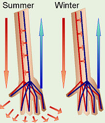
Countercurrent Heat Exchange
Much like a heat pump for your house or your refrigerator coils, your
cardiovascular system is also involved in countercurrent heating/cooling
of your body. Arteries and veins lying near each other in your extremities,
but flowing in opposite directions can absorb heat from each other as needed.
When your core temperature is too high, the arteries carry heat to the
extremities to be dissipated. As the blood returns via the veins, any
excess heat still in the blood is transferred to the arterial blood and sent
to the extremities, again. When your core temperature is too low, as the
blood flows out in the arteries to nourish the extremities, its heat is
transferred to the venous blood and sent back into the body to keep it
warm.
![]()

![]()