Respiratory System
2-D says, Heres where I am really different than you.
I dont have lungs, and I dont have gills, either. Along the sides of my
body are a series of holes called spiracles. We insects have a pair of
those on each abdominal segment, and usually on our thoracic segments as
well. Each of those spiracles opens into a tube called a
trachea, and all of those tracheal tubes are interconnected. Just like
your circulatory system is branched into smaller and smaller arteries, and
finally, the capillaries, similarly, my tracheal system is branched into
smaller and smaller tubes. Those tubes go directly to all my organs. That
means that outside air can come in a spiracle, then go down the tracheal
tubes until it goes straight to one of my organs. That means my blood doesnt
need to carry oxygen like your blood does, and I dont need hemoglobin like
you do. Because I dont have lungs, I dont need to breathe like you do,
but rather, the air in my tracheal system just gets to my organs by diffusion.
Some of my other insect cousins do have a chemical thats kind-of
like hemoglobin in their blood, except its called hemocyanin, it has a
copper atom in the center of its heme group (rather than the iron in the
center of your heme groups), and it gives their blood a sort of greenish-blue
color.
Background Information
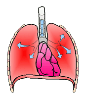
Breathing
(clipart edited from
Corel Presentations 8)
Each cell in an animals body must receive O2 and give off
CO2. This is easier for smaller organisms. In the vertebrates,
the blood carries O2 and CO2 to and from the cells, but
these gases must also be exchanged with the outside air or water. In insects,
the tracheal system takes air directly to the organs and O2 is
usually not carried in the blood. Mammals and some other vertebrates have
have lungs to exchange air. However, the lungs are ventilated differently
in different groups of vertebrates. For example, a frog opens its nostrils
and expands the floor of its mouth to draw air into its mouth. Then it
closes its nostrils and uses the floor of its mouth to push
O2 into its lungs. Mammals are unique in possessing a
diaphragm to pull O2 into the lungs. Even birds do
not have a diaphragm between their thorax and abdomen, so their liver and
heart are almost touching, and if their liver becomes enlarged for some
reason, it can rub on the birds heart. In mammals, as the diaphragm
contracts and the rib cage rises, a negative pressure is created in the
chest cavity causing the lungs to expand and air to be drawn in.
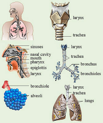
Respiratory System Diagrams
(clipart edited from Corel Presentations 8)
- Air first passes into the
nostrils where it is filtered by the nasal hairs and warmed
and humidified in the nasal cavity and sinuses.
- From there, the air passes through
the
pharynx,
which is shared with the digestive tract. Many students have trouble with
the pronunciation of this word. It is pronounced
fairinks ,
and you need to learn how to correctly pronounce it.
- Air next passes through the
larynx,
(pronounced as above, but with an l) also called the Adams apple or voice
box, and which contains the vocal cords. The vocal cords are under tension,
and a change in tension causes a change in pitch as air passes over them and
they vibrate. An inflammation of the larynx is called laryngitis.
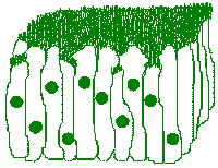
Pseudostratified Ciliated
Columnar Cells
- The larynx is situated at the top
end of the
trachea,
through which the air passes next. The trachea has rings of cartilage, like
the rings in a vacuum cleaner hose, for support. The lining of the trachea
is
pseudostratified ciliated columnar epithelium
which brushes debris up and out. This epithelial tissue is destroyed by
smoking, but can regenerate if the person stops smoking.
- The trachea divides at its bottom end
into two
bronchi
(sing. = bronchus), one to each lung. Recall that the mucus in the bronchi
serves to trap and coat dust particles so they dont scratch or infect the
delicate tissues in the lungs.
- The bronchi divide in the lungs
into smaller branches called bronchioles. In humans, the lungs are
not symmetrical because the heart, while located in the center of the chest
(thorax), leans slightly to the left. Thus the right lung has three lobes
(sections) and the left lung has two.
- The tiniest bronchioles branch to the
alveoli
(sing. = alveolus) which are tiny, multi-lobed air sacs made of simple
squamous cells. Having this thin wall enables air exchange with the
equally-thin-walled capillaries of the circulatory system. In order to
function properly, the alveoli must always stay moist. Special cells in the
alveoli secrete a substance called a surfactant which reduces the
surface tension of water, thereby enabling it to better coat the cells of the
alveoli to keep them moist and keep them from sticking to each other when the
person exhales. The ability to secrete this chemical doesnt develop until
around the eighth or ninth month of pregnancy, so there frequently is a
problem in premature babies with the lack of surfactant causing the alveoli
to stick together when the baby exhales. Then, when the baby inhales again,
the stuck alveolar cells tear away from their neighbors. Scar tissue forms
at these sites, thus the damage is permanent, and the persons lungs lose
some of their elasticity and ability to expand fully. A current hot area
of research is searching for a suitable replacement surfactant that could be
placed into the lungs of premature babies to prevent this damage.
The usual volume of air inhaled/exhaled in one breath is
called the tidal volume. The average tidal volume for an adult human
is around 500 mL of air. The maximum volume that can be exhaled during
forced breathing (as in the breathing machines people are given after
surgery) is called the vital capacity. For young adult male humans,
this amounts to around 4 to 5 L of air, and the average for females is
slightly lower.
As mentioned when we were discussing muscles, the diaphragm
is unique in that control of its operation can be either voluntary or
involuntary. Normally, control is involuntary, and we dont have to think
about breathing. The breathing center in the medulla of the brainstem
responds to O2 and CO2 content in the blood when
adjusting the breathing rate. We also have the ability, somewhat, to
control breathing voluntarily, and a classic example of this
is holding ones breath while swimming. I have heard that, in some kinds
of brain damage that affect the breathing center, the person may be able
to, at least partially, remember to consciously breathe while awake, perhaps
to the point of not needing mechanical help, but that person will need
a respirator to force air into his/her lungs at night while (s)he is
asleep.
Related to this, I have heard that it is physiologically
impossible for a person to hold his/her breath until (s)he suffocates (as
some young children will occasionally threaten to do).
Generally, as CO2 builds up, a point is reached where the person
just cant hold his/her breath any longer. If the person would pass out,
control would immediately return to involuntary, and (s)he would
automatically start breathing normally. Parents, do not give in to a child
who tries to do this to control you! I have seen advice that says to
ignore and not react to this type of behavior. One thing that might not
occur to you when you are upset with a childs behavior is that it would be
pretty difficult for the child to hold his/her breath while being gently,
lovingly tickled or if enticed into a conversation about some other,
interesting topic.
To get air to all the cells of the body, in mammals,
hemoglobin in the RBCs carries O2 to everywhere in the
body. However, hemoglobin has a greater affinity for carbon monoxide (CO),
and does not readily release it. Thus a victim of CO poisoning, is usually
put on supplemental oxygen to make sure the remaiming hemoglobin gets all it
can carry. Also, because of this, it takes a long time to recover from CO
poisoning. Some other organisms have hemocyanin in their blood (this
has Cu rather than Fe in a porphyrin ring). This is typical of many insects
with greenish or bluish blood. Most insects, however, do not depend on
their blood to take oxygen to their tissues, but rather, their tracheal
system allows air to go directly to the body organs.
Knowing CPR (cardiopulmonary resuscitation), or at
least mouth-to-mouth can prepare you to save someones life, and the
Heimlich maneuver (developed by a doctor here in Cincinnati) can help
save someones life if (s)he is choking. If you have never had CPR training,
you might wish to check with the Red Cross for their class schedule.
Respiratory System Disorders and Diseases:
Diseases and disorders of the respiratory tract include:
- Hiccups
- are spasms of the diaphragm thought to be caused by not enough
CO2 in the body. Thus, hiccups are frequently cured by
breathing into a paper bag.
- Rhinitis
- is an inflammation of the mucus membrane in the nose, due to a common
cold, allergies, etc.
- Pharyngitis
- is just a fancy name for a sore throat, which could be due to a viral
infection such as the common cold or flu or a bacteria infection such as
Streptococcus pyogenes (AKA strep throat).
- Laryngitis
- is an inflammation of the vocal cords in which the person partially
or totally loses his/her voice.
- Bronchitis
- is an inflammation of the bronchi, causing them to over-secrete
mucus, which in turn, causes coughing to get it up.
- Pneumonia
and
tuberculosis
- infect the lungs.
- Empyema
- is an infection, similar to pneumonia, in the chest cavity outside of
the lungs.
- Pleurisy
- is an infection of the pleural membranes lining the inside of the
chest cavity and coating the lungs. Normally these membranes are very
slippery, aiding in breathing, but when they become infected, they dont
slide over each other as well, and breathing becomes painful.
- Asthma
- is a reaction (often due to allergies) that causes constriction of the
bronchiole muscles,
thereby reducing the air passages, thus the amount of air that can get
to the alveoli. Interestingly, many of the treatments for asthma are
similar to treatments used for
hypoglycemia.
That and the fact that diabetics rarely also have asthma have led some
authors to suggest that asthma may be related to hypoglycemia, and that
a hypoglycemia diet may aid in alleviation of asthma symptoms.
- Emphysema
- is a progressive loss of elasticity in the lungs due to rupture of
some alveolar walls, coalescing of alveoli, and formation of scar
tissue.
- Lung cancer
- has been shown to be more common in people who smoke cigarettes and/or
who are constantly forced to inhale someone elses side stream smoke. A
number of pamphlets from the American Cancer Society and biology textbooks
have featured pictures that show what smoking can do to a persons lungs.
Typically, there is a photograph of a robust, healthy, pink lung next to a
photograph of a shrivled, diseased, blackened lung from a smoker.
Similarly, people who work around substances like asbestos fibers, coal
dust, flour dust, or dry, crumbled, dusty bird droppings for much of
their lives, frequently show signs of lung diseases caused by these
substances.
Copyright © 1996 by J. Stein Carter. All rights reserved.
This page has been accessed  times since 14 Mar 2001.