Survey of Invertebrate Phyla
Background Information on Kingdom Animalia:
Kingdom Animalia
includes about 35 phyla (depending on whose classification scheme is being
used). When many people think of animals, they only think of
mammals,
yet Class Mammalia is only one class in Subphylum Vertebrata, which is only
one subphylum within Phylum Chordata. We tend to lump all the other phyla
and other taxa within Vertebrata into a catch-all pseudo-group, the
invertebrates, but this is about 95% of all animals! There are more
species of insects than all other plants and animals together (The more
conservative estimates are that there are over one million species of just
insects.). Mammals are only a minuscule piece of the picture.
Animals live practically everywhere on this Earth. Insects,
alone, inhabit nearly all possible environments on Earth with the exception
of deep in the ocean, yet their close relatives, the crustaceans have
representatives that live there. Many other animals in many phyla live in
the ocean and in just about every terrestrial habitat.
In general, animal reproduction includes flagellated
sperm and a larger egg which join in fertilization to
form a
zygote.
This grows by
mitosis
to form a blastula (blasto = bud, sprout), an embryonic stage
that resembles a hollow ball, then on to other embryonic stages. From this
point, if the young resemble the adults, it is said that the embryo
grows into young, which grow into adults. If the young are very different
from the adults, it is said that the embryo grows into a larva
(larva = ghost, specter) which grows and metamorphoses into an
adult. Caterpillars and tadpoles are larvae, which, as is typical
of larvae, have different food, habitat, and appearance than the corresponding
adults. A larva undergoes metamorphosis (meta = between, with,
change; morpho = form; -sis = the act of) to become an
adult.
 Before discussing the various animal phyla, it is useful to introduce some of
the terminology which will be used to describe these animals. The back or
top side of an animal is its dorsal (dorso = back) side, and
its belly or bottom side is its ventral (vent(er) = underside,
belly) side. The head or front end is called the anterior
(ante = before) end, and the tail or back end is the posterior
(post = behind, after). Animals with radial symmetry
(radia = spoke, radius; sym = with, together, metr,
-metry = measure, measurement) do have distinct top and bottom sides,
but have no distinct left and right. Starfish, jellyfish, and sea anemones
are examples of animals with radial symmetry. Animals with bilateral
symmetry (bi = two; later = side) do have distinct left
and right sides, and most animals with which we are familiar, such as
earthworms, ladybugs, and dogs, have bilateral symmetry.
Before discussing the various animal phyla, it is useful to introduce some of
the terminology which will be used to describe these animals. The back or
top side of an animal is its dorsal (dorso = back) side, and
its belly or bottom side is its ventral (vent(er) = underside,
belly) side. The head or front end is called the anterior
(ante = before) end, and the tail or back end is the posterior
(post = behind, after). Animals with radial symmetry
(radia = spoke, radius; sym = with, together, metr,
-metry = measure, measurement) do have distinct top and bottom sides,
but have no distinct left and right. Starfish, jellyfish, and sea anemones
are examples of animals with radial symmetry. Animals with bilateral
symmetry (bi = two; later = side) do have distinct left
and right sides, and most animals with which we are familiar, such as
earthworms, ladybugs, and dogs, have bilateral symmetry.
The body of an animal is made up of several layers of tissue.
Ectoderm (ecto = out, outer, outside; derm = skin) is
the outer layer of tissue. The epidermis (epi = upon, over),
or skin, other outer layers, and the nervous system in vertebrates (not in
all animals) are formed from ectoderm tissue.
Mesoderm (meso = middle) is the middle layers of tissue.
Mesoderm forms the muscles and most other internal organs. Endoderm
(endo = within, inner) forms the inner layers, including the lining
of the digestive tract in all animals and the liver and lungs in vertebrates.
Often animals have a space in their bodies between several of these layers.
Animal groups, like flatworms, with no such space are referred to as
acoelomates. Animals, such as roundworms, which have a space between
the mesoderm and the endoderm are called pseudocoelomates, and the
space is called a pseudocoelom. Animals, including earthworms,
insects, and humans, which have the space in between several of the mesoderm
layers are called coelomates, and the space is called a
coelom.
Background Information on Invertebrates:
In this lab, you will become familiar with the characteristics
of a variety of invertebrate phyla.
While Phyla Annelida, Phylum Arthropoda, and Phylum Chordata Subphylum
Vertebrata will be covered in subsequent labs,
this lab will survey some of the other invertebrate animal phyla. Some of
the characteristics of each phylum are listed below. Depending on what
specimens and slides and how much time are available, your instructor may
choose to focus on a more-limited subset of these organisms. A whole course
could be spent on comparative invertebrate anatomy, so this lab designed to
just skim the surface and provide an introduction to these organisms.
As time allows, examine
any microscope slides, plastic mounts, preserved, dead, and/or live
specimens of these organisms which are available. Take notes on everything
you see, draw pictures of each, and label all the parts, systems, and organs
that are visible. Include labeled drawings of external and internal anatomy
for as many of these animals as you are able to view.
Phylum Porifera
(pori = pore, small opening; fer = to bear,
carry)
The members of this phylum are commonly known as the sponges.
This is the most primitive group of animals, and it is thought that they
evolved separately from all other animal groups. Interestingly, they share
many characteristics in common with some forms of colonial protozoans.
Sponges have no symmetry.
The body consists of two layers of cells: the outer dermal
epithelium and the inner gastral epithelium which lines the
central cavity. Between these two layers of cells is a jelly-like
substance, and embedded in that, a skeletal framework, often
consisting of spicules of varying chemical composition.
There are many pores in the body wall, hence the group name,
Porifera. In general, water, along with potential food, flows into these
pores, and out through the main ostium or excurrent opening.
Sponges are
hermaphroditic,
that is, they have both sexes in one body. This term is derived from the
myth in which Hermes (Mercury) and Aphrodite (Venus) got together and had a
son, who they named Hermaphroditus, who while swimming one day, somehow
became united in one body with a water nymph. Sponges can reproduce sexually
via eggs and sperm (which are just released into the water), with the
resulting zygote giving rise to a multicellular, free-swimming larva.
Sponges can also reproduce asexually by budding (a piece breaks/pinches off
and forms a new colony of sponge. As adults, they are sessile (attached to
one place).
Class Calcarea

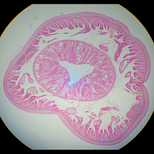
Grantia sp., longitudinal section (too big to fit under low power all at once)
This photograph is a longitudinal section (l.s.) through a
small sponge called Grantia, and exhibits the body structure typical
of sponges. (This slide is not in your slide box, and we will not be
examining it during lab, but it is included here so that you can better
understand how a sponges body is put together.)
Not visible in this microscope slide is the excurrent pore,
the opening at the top of the body of this sponge.
Below is a close-up view of a portion of the upper left corner
of the above photograph.

Enlarged view of a portion of Grantia sp.
The central cavity in the body is called the gastral
cavity. Notice how the body wall zigzags back and forth forming a number
of canals. Incurrent canals open towards the outside, and are, thus,
lined with dermal epithelial cells. Radial canals open toward the
gastral cavity, and are lined with gastral epithelium. Notice that there are
many small pores that connect the incurrent and radial canals. Water
(carrying food, etc.) flows into the incurrent canals, through the pores
into the radial canals, then into the gastral cavity. The cells of the
sponge filter out, eat, and process the food, then excrete the wastes into
the water in the gastral cavity. From there, the water is released via the
excurrent canal at the top of the sponge.
Class Demospongiae
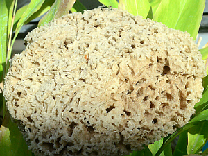 Spongia officinalis Commercial Sponge
The body plan of these large sponges is arranged similarly to that seen in
Grantia, but much more complex, consisting of a very complicated
network of interconnecting canals. These sponges are collected from the
ocean, then left to dry, so all the cells died long ago, and what is left
is the skeleton of the sponge, made of fibers of a chemical called
spongin, which is a type of protein and is chemically similar to
the collagen in our bodies. (Some other types of sponges also contain a
skeletal structure called spicules which are spiky and glasslike.)
Examine the skeleton of a
commercial sponge and note the complex network of canals of which it is
comprised. Also, observe any available plastic mounts of sponges.
(A side note: the cellulose sponges that we all use, now,
were designed to mimic the looks and water-absorbing ability of real
sponge skeletons.)
Phylum Coelenterata or Cnidaria
(coelo = hollow; entero = intestine, gut;
cnida = stinging nettle)
This phylum includes animals like jellyfish (Class Scyphozoa),
coral and sea anemones (Class Anthozoa [anthe = flower;
zoa = animal]), all of which are marine, plus Hydra, a
freshwater genus (Class Hydrozoa).
These animals have radial symmetry.
The body wall consists of two layers of tissue:
ectoderm/epidermis on the outside, and endoderm/gastrodermis lining the
gastrovascular cavity, with a non-living, jelly-like mesoglea in
between them.
They have a digestive cavity with one opening, which thus is
called a gastrovascular cavity (gastro = stomach; vascul
= a little vessel). The name Coelenterata refers to the gastrovascular
cavity, so named because it serves the functions of both digestion
(similar to our digestive system) and distribution (similar to our
circulatory system) of the food. Also, it has one opening which serves the
functions of both a mouth and an anus (food, etc., in, wastes
out).
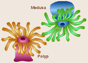 While, overall, the bodies of these animals are similar, are all the same
basic shape, they can take one of two forms, and thus, the bodies of
cnidarians are described in one of two ways, depending on
whether the opening of the gastrovascular cavity is dorsal (up) or ventral
(down).
Animals like Hydra and sea anemones, which are sessile
(anchored to the substrate and not moving much, if any) and have the
opening (mouth) and surrounding tentacles at the top of their bodies have a
body shape called a polyp (polyp = many footed).
Jellyfish, which float (or are free-swimming) and have the opening and
surrounding tentacles at the bottom, have a body form called a
medusa (medusa = a jellyfish).
While, overall, the bodies of these animals are similar, are all the same
basic shape, they can take one of two forms, and thus, the bodies of
cnidarians are described in one of two ways, depending on
whether the opening of the gastrovascular cavity is dorsal (up) or ventral
(down).
Animals like Hydra and sea anemones, which are sessile
(anchored to the substrate and not moving much, if any) and have the
opening (mouth) and surrounding tentacles at the top of their bodies have a
body shape called a polyp (polyp = many footed).
Jellyfish, which float (or are free-swimming) and have the opening and
surrounding tentacles at the bottom, have a body form called a
medusa (medusa = a jellyfish).
This phylum is noted for the specialized stinging cells
(cnidocytes or nematocytes) which are found only in this
phylum and are located on the tentacles. The combination of tentacles and
cnidocytes is used to capture and paralyze prey. The toxins in some jellyfish
cnidocytes are irritating or toxic to humans.
Cnidarians have both sexual and asexual forms of reproduction.
Hydra, for example, will periodically develop lumps on the sides of
their bodies that are either testes or ovaries. These make and release
sperm or eggs, which then join to form a zygote, which eventually grows into
a new hydra. Hydra also reproduce asexually by a process known as
budding in which a new, small Hydra begins to grow from the
side of a large Hydra, eventually pinching off when it is fully
formed.
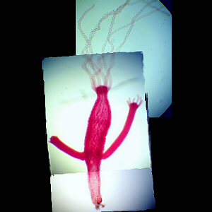
Class Hydrozoa

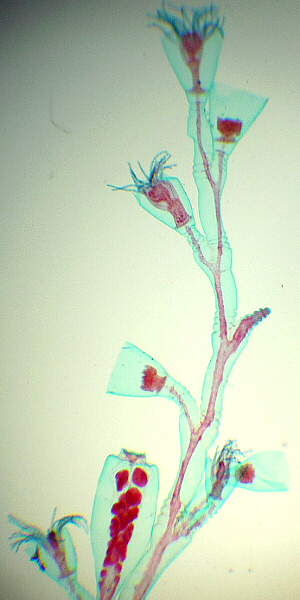 Obelia sp. have both polyp and medusa forms in different stages of
their life cycle.
(This slide is not in your slide box, and we will not be examining it during lab,
but it is included here so that you can compare the polyp and medusa forms.)
Obelia sp. have both polyp and medusa forms in different stages of
their life cycle.
(This slide is not in your slide box, and we will not be examining it during lab,
but it is included here so that you can compare the polyp and medusa forms.)
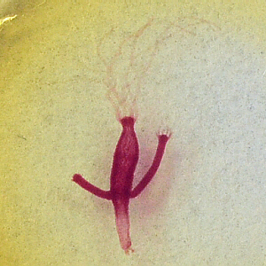
Hydra sp. Hydra (all are polyp form)
Several Live Hydra
Live Hydra
Hydra is the only common fresh-water coelenterate. They may
be found in local streams, but due to their small size, probably wouldnt be
noticed unless someone was specifically looking for them.
The body of a hydra consists of a stalk, the bottom end
of which is called the base or basal disc. The top end
consists of the tentacles, and the hypostome (the area around
the mouth), and the mouth itself (or oral opening).
Hydra can move either by gliding on their bases or by turning
somersaults.
Especially on the tentacles, hydra bear cnidocytes or
stinging cells (hence the name of this phylum). These cells discharge to
paralize and capture prey.
Hydra with Buds, wm, Actual View
Hydra with Buds, wm, Microscope View
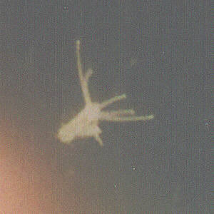
Live Hydra with Bud (Actual Color)
Hydra can reproduce asexually by budding, in which a new hydra grows on
the side of an older one (as in these photos), then eventually pinches
off.
View the
prepared slide of a hydra budding (Carolina #Z615). Draw your budding hydra,
and label all the body parts listed above.
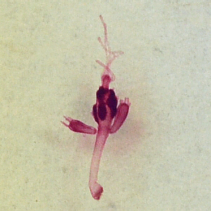
Hydra with Spermaries, wm, Actual View
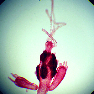
Hydra with Spermaries, wm, Microscope View
In sexual reproduction, hydra can form testes (a smaller lump thats
nearer the oral opening and sticks out more, as above), and/or ovaries (a
larger lump thats nearer the base and sticks out less, as below).
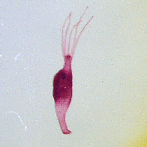
Hydra with Ovaries, wm, Actual View
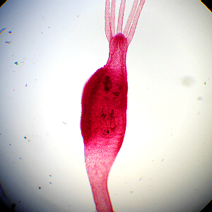
Hydra with Ovaries, wm, Microscope View
View the
prepared slides of hydra with spermaries (Carolina #E28) and with
ovaries (Carolina #Z640). Draw each hydra and label all its parts.

Hydra, xs, Microscope View
The body wall of a hydra consists of two layers of cells, the outer ectoderm
and the inner endoderm. Note that, unlike our bodies, they do not
also have a mesoderm layer. Also unlike our bodies, their ectoderm and
endoderm layers are each only one cell thick. There is a jellylike substance
called mesoglea between the ecto- and endoderm. The ectoderm is
comprised of epitheliomuscular cells which serve both as a body covering
and contain muscle fibrils for movement. Periodically interspersed among
the epitheliomuscular cells, and especially numerous on the tentacles, are a
few cnidocytes or stinging cells, which can only function once, then
must be replaced.
The most prevalent cells in the endoderm are nutritiomuscular cells,
which aid in intake of food and body movement. Some bear flagella which
help to circulate the contents of the gastrovascular cavity. In the
endoderm there are also some gland cells which secrete digestive
enzymes into the gastrovascular cavity.
(This slide is not in your slide box, and we will not be examining it during
lab, but it is included here so that you can better understand how a hydras
body is put together.)
Class Scyphozoa
While some kinds of jellyfish are hydrozoans, many types of
jellyfish are scyphozoans. All members of this class are of the medusa
form.
(This group is mentioned just because it fits in, here, but we will not be
examining them.)
Class Anthozoa
Members of this class are of the polyp form. This group
includes sea anemones and coral. Corals are colonial, secreting
protective structures in which they live. These coral reefs area often very
colorful.
(This group is mentioned just because it fits in, here, but we will not be
examining them.)
Phylum Platyhelminthes
(platy = broad, flat; helminth = a worm)
Members of this phylum are collectively known as the flatworms
because their bodies are flat and ribbonlike. They have bilateral symmetry,
but are acoelomates (have no coelom). They have a gastrovascular
cavity with one opening. Most have no respiratory structures and breathe
through their skin. Some of these are free-living (aquatic), and others
are parasitic.
Class Turbellaria
This class includes a number of genera and species which are
collectively referred to as Planaria, as well as other, closely-related
organisms. They are all free-living, and there are both fresh- and
saltwater forms (all are aquatic). Planaria typically are found in
freshwater streams, including those here on campus.
Members of this class have a gastrovascular cavity with one opening.
They have bilateral symmetry they have specific dorsal and ventral
sides, anterior and posterior ends, and right and left sides.
This is the first group of animals in which all three tissue
layers are present. Externally, there is a layer of epidermis
or ectoderm, the ventral surface of which bears cilia. These enable a
gliding motion.
Internally, there is a layer called the endoderm or gastric epithelium or
gastrodermis which lines the gastrovascular cavity. This
is the first group of animals in which a layer of mesoderm is found
in between those two layers. This consists of layers of muscles
just under the epidermis, as well as a layer of mesenchyme tissue,
which takes up most of the space in the body.
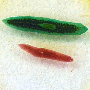
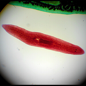
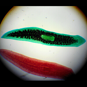
Planaria, Actual View of Slide
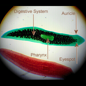
Labeled Planarian
The first picture is an actual photo of the microscope slide of two stained
planarians, the
others are microscopic views. These are stained whole mounts. The
green-stained one shows the extent of the digestive system. Note the
pharynx and the light-sensitive eyespots.
The pigment within the eyespots in the head region
is light-sensitive, but these eyes do not see images like our eyes
do. The lateral extensions in the head region are called auricles.
In the middle of body, on the ventral side there is a
pharynx which may be extended or retracted. This serves the dual
purposes of taking in food and elimination of waste. Since, once again,
there is only one opening to the body cavity (digestive system), that is,
again, referred to as a gastrovascular cavity.
Planarian
Planarian Stained to See Digestive System
Planaria are reported to feed on algae and the dead bodies of snails and
other aquatic organisms. When kept in captivity, they are often fed bits of
liver.
Planarians have an excretory system made up of a network of
tubules with flame cells at the ends (this system may be
visible on specially-stained, prepared slides) to eliminate water,
CO2, and nitrogenous wastes.
They have a well-developed nervous system to coordinate
feeding, movement, etc. This is a ladder-like nervous system
with nerve cords running down each side and rungs connecting them. There
are a number of ganglia, including a larger ganglion (brain) in the head
region. There are also sensory structures, including the eyes.
Planaria have sexual reproduction. They are
hermaphroditic (have both male and female gonads/genitalia) and
cross-fertilize when they mate with another. They can also reproduce
asexually by pinching apart, and each half regrowing the missing
portions. A common lab demonstration that is done is to cut apart a
planarian in various regions of the body to observe its regenerative
capabilities, and that is a hot research topic because, if we could
understand how they are able to regenerate missing parts, it is reasoned that
perhaps that would give us a way to turn those genes on in humans.
(Other experiments have also been done to study their learning
ability.)
View the prepared slide
of a whole mount (wm) of stained planarians (Carolina #Z915) under low power
(the slide is too thick and will crack if you attempt to use higher powers
of magnification. Draw and label all body parts. Notice that the
gastrovascular cavity has one anterior branch and a pair of posterior
branches (for a total of three branches/parts).
Observe any plastic mounts of planaria.
If live planaria are available, watch one move. If available, try adding some
food to the water, and see if the pharynx is extended.
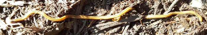
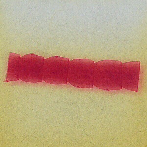
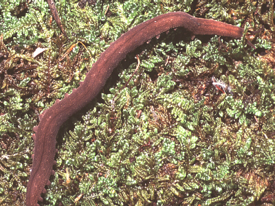
Bipalium kewense Land Planarian (© 2016 by Eric Stein)
Heres a planarian relative that, if you live here in the US,
you may not see. (It certainly is not one I was told about back when I was
in school!) These are sometimes called hammerhead slug or hammerhead
worm, but they are not slugs, and they are not related to earthworms.
They are thought to be from somewhere in Indo-China, and are not native to
the US, but were first seen here in 1901, in greenhouses. In greenhouses,
they live in the moist environment in potted plants, and thus, can be spread
when the potted plants are shipped to somewhere else. In some warmer areas
of the US, they have become established outdoors, while elsewhere, they may
be present seasonally (due to planting new plants in whose pots they were
living). This individual was photographed in June 2016 in my brothers
yard in the Smoky Mountains in Tenn.
Our native planarians are typically in the vacinity of 1 cm long and aquatic.
These planarian-relatives are terrestrial, and can grow to over 10 in (25 cm)
long. True to some of their common names, their head (to the left in this
photo) is hammerhead-shaped.
Like our local, aquatic planaria, they may have eyespots (again, not true
eyes) on their head.
Also, as described above, they both feed and eliminate wastes via a pharynx
midway down their body. Like our local planaria, they are also carnivores,
with earthworms being a
favorite food (people who own earthworm farms and are raising earthworms for
money dont like them), though according to one Web page, the will also eat
slugs and some types of insect larvae.
Like our local planarians, theyre really good at regeneration. While they
do reproduce sexually, apparently reproduction via the end of the tail
pinching off is more common... and, just like the locals, if someone cuts
one into several pieces, each piece can grow into a new Land Planarian.
They move by secreting a mucus trail, along which they can glide by using
cilia.
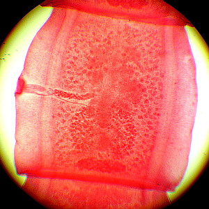
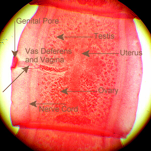
Class Trematoda
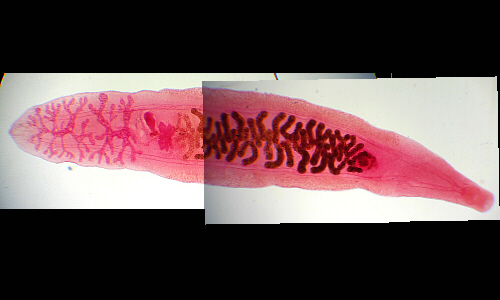
Clonorchis sinensis Chinese Liver Fluke
The members of this class are known as flukes. They are
parasites in the liver, lungs, intestines, or blood of various animals, and
have complex life cycles needing specific hosts. One
of the most frequently-studied is the Sheep Liver Fluke, Fasciola
hepatica, a parasite in the liver of sheep. (Note that the prepared
slide, above, is of a different species. This slide is not in your slide
box, and we will not be examining it during lab, but it is included here so
that you can better understand what a fluke looks like.)
The Sheep Liver Fluke has an anterior sucker to feed
on the liver of its host. About a fifth of the way down the body from the
anterior end is the ventral sucker which is used to attach to host
tissue. Midway between these two suckers is the genital pore. At
the posterior end is an excretory pore, the opening of the excretory
system.
The digestive tract has two lateral branches running
posteriorly down each side. Flukes are hermaphroditic. The flame
cells of the excretory system are very difficult to see without a
specially-stained prepared slide.
Interestingly, flukes have no epidermis (ectoderm) layer.
Rather, the mesoderm muscle tissue is directly covered by a
non-living cuticle.
Several species in genus Schistosoma are known as
blood flukes and are parasites whose life cycle involves both humans and
snails. A human who is infected with them is said to have schistosomiasis.
Schistosomiasis ranks second, after malaria, in terms of the number of people
(many in third-world countries) who are infected. The flukes parasitize blood
vessels in the mesentaries (the membranes which surround the intestines).
Members of genus Schistosoma are unusual, compared with other flatworms
and even other trematodes, because they are dioecious they have
distinct, separate male and female flukes (remember other flatworms are
hermaphroditic). Eggs are eliminated in either the persons urine or feces,
depending on the species of Schistosoma involved, and then the larval
stage must parasitize a snail host. Then, humans become infected when they
wade in snail-containing water in tropical areas where this fluke occurs,
and a next-larval-stage organism burrows right through their skin. Just as
a major component of malaria prevention is based on treating water supplies
to eliminate mosquito larvae, so also, a major component of prevention of
schistosomiasis is treating water bodies to eliminate snails. There is
also a drug that may be given to humans who are already infected, and often,
if in a particular village, a large number of children are showing symptoms,
the simplest thing to do is to just treat everyone in the village, which
also helps to eliminate re-contamination of the local water supply. Also,
simple preventative measures can be taken: just as the use of bed nets
eliminates mosquitoes ability to get to humans (children) to spread malaria,
so also educating people (children) to avoid going in the water (no swimming)
can cut down on the number of people infected with this parasite.
Observe any plastic mounts
of flukes that are available.
Class Cestoda
The members of this group are known as tapeworms due to their
shape, and are all parasitic. Their bodies are made of a number of segments
called proglottids, each of which is pretty much on its own and
separate/independent from the rest. These proglottids dont need a digestive
system because they absorb pre-digested food from the hosts digestive tract
and dont need a respiratory system because they absorb dissolved air
(O2), again, from the fluids in the hosts digestive tract.
Thus, these segments are mostly reproductive system (imagine each segment as
a very prolific female!). The segments break off and are passed out with
the hosts feces. Other animals who accidentally ingest some of the
infected feces (cattle eating grass near where other cattle have defecated)
can acquire the parasite. Tapeworms can grow very large (long) and absorb
so much food from their hosts digestive tract that they cause nutritional
deficiencies.
One commonly-studied example is the Dog Tapeworm, Taenia pisiformis.
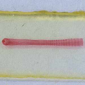
Anterior Segments and Scolex
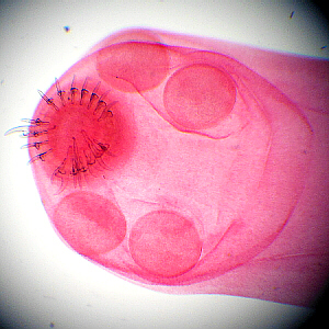
Close-up of Scolex

Labeled Scolex
The first segment of the body is called the scolex, and the rest are
called proglottids. The proglottids closest to the scolex are the
newest, youngest ones, and those farther away are older. The purpose of the
scolex is attachment. It has a ring of hooks plus four suckers to attach to
the wall of its hosts intestine.
Mature Proglottids
Close-up of Mature Proglottid
Labeled Proglottid
While these structures may not be easily distinguishable, in the proglottids,
a nerve cord and an excretory tube run along each side. The excretory
system has one cross-connection posteriorly in each segment.
Neither the organism as a whole, nor the individual segments has any kind of
mouth or digestive tract. Mature proglottids contain essentially no other
body organs than (or lose all but) a reproductive
system (each segment has its own) with a lateral genital pore. They
are hermaphroditic, and may self-fertilize, or may copulation with another
proglottid when the chain folds back on itself bringing proglottids into
contact with others.
Once fertilization occurs, as the eggs
develop and mature, all
other systems disintegrate, so the older proglottids are, essentially, just
egg sacs. Those at the end of the chain break off and are passed out with
the hosts feces. Those eggs may, then, be ingested by another host animal
who eats, for example, grass from near where those feces were deposited. If
dog tapeworm eggs are ingested by rabbits, they hatch and burrow into the
rabbits muscle, and form a cyst. If that rabbit is subsequently eaten by a
dog, the tapeworm comes out of the cyst form, attaches to the dogs
intestine, and grows there.
View the prepared slide of
a tapeworm (Taenia sp.) scolex (Carolina #Z971) and the prepared
slide of tapeworm (Taenia sp. mature proglottids (Carolina # Z973b).
Draw what you see and label all body parts that are visible.
Also, view (and draw) any plastic mounts that are available.
Phylum Nematoda
(nema, nemato = a thread)
Members of this phylum are called roundworms because their
bodies are cylindrical. Some are free-living in aquatic habitats (including
damp soil), while others are parasitic. They have bilateral symmetry.
For the first time, here, were seeing animals
with a tube-within-a-tube body plan, in that they have both a mouth
and an anus, and food travels through the digestive system from one
end to the other. They have all three layers of tissue: ectoderm,
mesoderm, and endoderm with a pseudocoelom (false
coelom), a cavity between the meso- and endoderm (a
true coelom is between meso- and more mesoderm). All muscle fibers
in their bodies run longitudinally, so only they can only do thrashing
movements. There are free-living (in water and in moist soil) and parasitic
forms which range in size from microscopic to several feet in length.
They have sexual reproduction with definite males and females.
Class Secernentea
One commonly-studied roundworm is Ascaris lumbricoides,
a parasite of pigs. The smaller male has a curled posterior end and the
larger female is straight. They are unsegmented and have no conspicuous
appendages or sense organs.
On the female, the posterior end is more pointed than the
anterior, and has a slit-like anus near the tip on the ventral side. The
anterior end has three lips surrounding the mouth. About a fourth of the
way down, on the ventral surface, is a genital pore. There is a faint
lateral line on each side, indicating the lateral excretory canal.
There are also less-distinct dorsal and ventral lines, indicative of the
dorsal and ventral nerve cords.
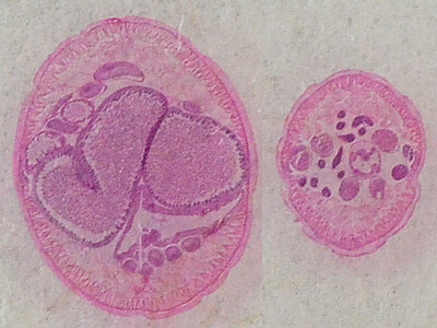
Ascaris lumbricoides, x.s., Intestinal Region
Ascaris lumbricoides is a roundworm that is parasitic on pigs. This
is a cross-section through the female (left) and male (right) showing the
uterus full of eggs/embryos in the female and other internal organs in both.
Ascaris lumbricoides, x.s., Esophageal Region
This slide shows the internal organs in the region of the esophagus.
(These two slides are not in your slide box, and we will not be examining
them during lab, but they are included here so that you can better
understand what roundworms look like.)
Class Adenophorea
Male and Female Trichinella Worms, w.m.
Trichinella spiralis (trichin = a hair; -ella = small) is
a parasitic roundworm that infects muscle tissue in mammals that happen to
consume some of them, and seems to be especially prevalent in pigs. The
worms form cysts in the hosts muscle tissue. The infection is called
trichinosis (trichin = a hair; -osis = diseased
condition or state), and can be transferred to humans via consumption of
(especially) pork that has not been cooked thoroughly enough to kill the
parasites that have encysted in the pigs muscle tissue.
Thorough cooking at high temperatures kills the parasites, but
microwaving often does not get the center of a piece of meat hot enough for a
long enough time to be effective.
Because of this, while it is OK to reheat pork in a microwave, it
should never be cooked that way for the first time. If a person ingests
living Trichinella in undercooked meat, in the persons intestines,
the parasites will develop from larvae into adults and reproduce. Mated
females will bore into the persons intestinal muscles, where they will
produce a new generation of larvae. These new larvae, in turn, either travel
through the persons lymph system or burrow through his/her body tissue to
reach his/her muscles and other tissue where they will live.
Encysted Parasites in Muscle Tissue

Close-up of Worm in Cyst in Muscle
Parasitic roundworms also include pinworm and hookworm.
School children frequently contract pinworm from doorknobs, etc. at school
because infected classmates dont wash their hands after wiping feces from
their anal areas.
Pinworms live in the rectum and lay their eggs around the anus, thus one
main sign of a pinworm infection is when a child frequently scratches
his/her anal area. A classic test for pinworm infection is to touch a piece
of scotch tape to the childs anal area, followed by microscopic examination
of the tape to check for the presence of pinworm eggs.
Many roundworms are free-living, but need to live in an
aquatic environment of some sort, which can include damp soil. Many soil
nematodes are beneficial and are important decomposers, but there are some
which can be found in garden soil that damage desirable plants.
View the prepared slide of
Trichinella spiralis in muscle (Carolina #PS2430). Find a good
looking encysted parasite to draw and label.
Phylum Rotifera
(rota = wheel, revolve; fer = to bear, carry)
These organisms are frequently seen when examining pond water
under a microscope. They may be identified by their characteristic rings of
cilia around the mouth which, when beating, look like rotating wheels, hence
the group name. These direct food into the mouth.
One of the most commonly-seen forms has a telescoping body and
a forked foot at the posterior end, but other configurations also occur.
They are all microscopic, but multicellular, and there are both fresh- and
saltwater-dwellers.
They also have a pseudocoelom like the nematodes. They have
a tube-within-a-tube body plan with a mouth and an anus. They have an
excretory system similar to flatworms. Frequently, they reproduce
parthenogenetically (partheno = a virgin, without fertilization),
that is, the females produce more females without fertilization by males.
While smaller and not as common, males do exist.
(This group is mentioned just because it fits in, here, and this may be
something you might see in pond water, but we will not be examining them
in this lab.)
Phylum Annelida
Taxonomically, Phylum Annelida would fit here. More
information on them can be found on the
earthworm Web page.
Phylum Onchyohora
(onycho = a claw, nail; phora, fer = to bear, carry)
Macroperipatus torquatus from Trinidad © M. K. Busching
Phylum Onchyophora includes the velvet worms (also called walking worms) in
genera Peripatus (peri = around, -patus = a walk, path)
and Macroperipatus (macro = large, long). This photo of
Macroperipatus torquatus, a larger species from
Trinidad, was taken by Milan Busching, Curator, Emeritus, of Invertebrates at the
Cincinnati Zoo.
This phylum is of interest
because its members share some traits in common with the Annelida, and other
traits in common with the Arthropoda, and thus, many biologists feel that,
evolutionarily, this phylum is midway between and may be an evolutionary
link between those two phyla. They do also have unique traits of their
own that are not shared with either the annelids or the arthropods.
One prevalent theory is that Onychophora is thought to have evolved from a
marine worm-type ancestor, with the parapods evolving into unsegmented legs.
Onychophorans have one pair of these unjointed, cone-shaped appendages per segment, and
those appendages are muscular and can move, but are not jointed as in the
arthropods.
This theory regards onychophorans as a sort-of missing link on the way to
the evolution of insects (Other biologists believe that arthropods arose
directly from annelids through some jointed-legged ancestor.).
Onychophorans walking legs have a pair of claws at the tips. At the
anterior end of their bodies is a pair of antennae plus a pair of
oral papillae, and in the mouth, a pair of jaws, all three of which
are modified appendages, similar to the condition in arthropods.
Onychophorans are predatory and shoot sticky, mucus/saliva to tangle their
prey.
They have a tubular digestive system.
They are bisexual (have males and females) and are live-bearing (viviparous).
There is a pair of simple eyes on the head.
The nervous system is simple, consisting of a brain and paired, ventral
nerve cords without distinct segmental ganglia.
Their excretory system consists of paired, segmental nephridia like annelids,
and locations of these corresponds to the locations of the legs.
They also share the presence and structure of their cuticle, their ciliated
reproductive tract, and their musculature with Phylum Annelida.
However, their respiratory system of
tracheal tubes which distribute oxygen directly to the various organs, paired
appendages with claws at the tips, open circulatory system,
and chitinous exoskeleton which must periodically be molted are
shared with Phylum Arthropoda.
View the plastic mount
of Peripatus.
Phylum Arthropoda
Taxonomically, Phylum Arthropoda would fit here. More information
on them can be found on the
crayfish Web page,
the
eyelash mite Web page,
and the
insect Web page.
Phylum Mollusca
(mollusc = soft)
There are several classes of mollusks:
- Class Polyplacophora (poly = many; plakos = flat
plate, tablet; phora = bear, carry) which includes the chitons
- Class Gastropoda (gastro = stomach; poda = foot)
which includes snails and slugs
- Class Bivalvia or Pelecypodia (bi = two) which includes
oysters and clams
- Class Cephalopoda (cephalo = head) which includes
octopus (octa = eight), squid, and chambered nautilus
Mollusks are soft-bodied and have a true coelom. Many have
shells made primarily of calcium carbonate: chitons have a shell made of
eight plates, snails have one, spiraled shell, clams have a shell composed
of two, hinged halves, squid have small internal shells, and the chambered
nautilus has a chambered shell (spiraling in living species but straight in
many fossil forms).
The general body structure includes a ventral, muscular
structure, the foot, which is involved in locomotion. Dorsal to
that is the visceral mass, including the internal organs. Covering
part to much of the body is the mantle, which often secretes a
calcium carbonate shell. In the anterior portion of the digestive
tract is a radula (radul = a scraper), a rasping organ used to
obtain food. Most dont show much sign of segmentation, with the exception
of the chitons. They have bilateral symmetry. Aquatic forms
have gills for exchange of oxygen and carbon dioxide.
While snails and slugs have a radula (radul = a
scraper), a rasping organ in their mouths to scrape bits of food into
their mouth, most bivalves use their gills to filter small food particles,
which are then directed toward their mouths. Cephalopods have beaklike jaws
for biting and crushing their prey, and unlike members of the other classes,
tend to be fast-moving carnivores.
Many mollusks have good eyes (snail, octopus, squid).
Octopuses are famous for their well-developed nervous systems and brains
(necessary to process the information from their sophisticated eyes), and
they are known to be very intelligent.
Most mollusks have males and females, while certain kinds of
garden snails are hermaphroditic.
Examine any plastic mounts
that are available. Additionally, since many of these are used as food
by humans, samples may be available from the seafood section of a local
grocery store.
Class Gastropoda
(gastro = stomach; poda = foot)
This class includes the snails and slugs. Some are marine,
some inhabit freshwater, and some are terrestrial.
Snail Eye
At their anterior (head) end, snails and slugs have two pairs of retractable
tentacles with eyes at the tips of the longest pair. Because snails
have a one-piece shell, they are called univalves. As snails and
slugs move,
they secrete a layer of mucus upon which they glide (hence the slug trails
seen on sidewalks). Some types of snails are hermaphroditic.
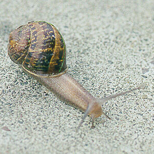
Snail
Snails and slugs have a rasping radula that helps them ingest
their food.
The visceral mass forms a
dorsal hump, covered by the mantle which secretes the
shell. The digestive system (and other internal organs) is modified
to account for the position of the visceral mass, so it goes up into the
portion of the body held within the shell, around, and back out, again,
somewhat anteriorly. While marine forms have gills, terrestrial snails have
the inner surface of the mantle cavity modified to form
lungs, instead.
Out in California, there is a species of
slug called a Banana Slug. These are large approaching the size of a small
banana and bright yellow in color.
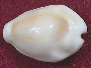
Cowrie Shell, Top Side
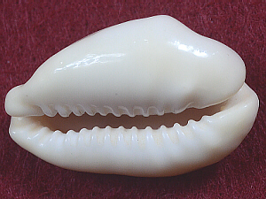
Cowrie Shell, Bottom Side
Cowries are another type of gastropod. They are marine, and like snails,
their bodies extend inside their shells. Humans from many cultures have
found their porcelain-like shells to be attractive.
Class Pelecypoda
(pelecy = hatchet, axe this name is due to the
wedge-shaped foot)
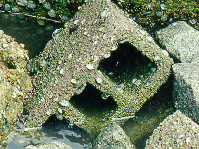
Mussels at Low Tide
This class includes mussels (note spelling), clams, scallops,
oysters, and similar organisms.
These have two-part shells, so are called bivalves. The two
halves are held together by a tough ligament in the dorsal region. Due to
the way the mantle secretes the shell as it adds to the shell, the shell has
growth rings. The widest side-to-side, oldest part of the shell is called
the umbo. Looking from the side, the shell is not symmetrical, and
the umbo is displaced toward the anterior end, while the other end is
posterior. The two valves of the shell open ventrally, and the foot
extends from there.
Bivalve Shells Used for Decorative Craft
The two halves of the shell are held shut by the large,
anterior and posterior adductor muscles, located dorsally. Just
inside the shell is the mantle, and under the mantle, the wedge-shaped foot
and the visceral mass. Modifications of the mantle create an internal
current with the mantle forming incurrent and excurrent canals.
This current brings in food and carries away waste. There are also two
pairs of gills, sheet-like structures attached to the dorsal part of the
body on either side.
Along the anterodorsal margin of the foot are two pairs of
flaplike labial palps, and the mouth lies midway between the
palps.
They have an open circulatory system. In the visceral mass,
the coelom is modified as a pericardial cavity. The heart is in that
cavity, surrounding the intestine. It pumps blood both anteriorly and
posteriorly into arteries, many of which open into blood sinuses rather
than capillaries. While passing through the gills, the blood absorbs
oxygen.
A nephridium lies near the pericardial cavity, and absorbs
nitrogenous waste from the pericardial fluid and blood, then
discharges that into the water flowing through the inside of the shell.
The nervous system consists of three pairs of ganglia: the
head ganglia near the anterior adductor muscle, the visceral ganglia near
the posterior adductor muscle, and the foot ganglia in the foot. These
ganglia are joined by paired connectives.
These organisms are bisexual (have separate male and female
organisms). There are both freshwater and marine species.
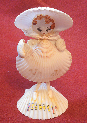
Humans enjoy using mollusk shells in craft projects. The
doll in this photo
is composed of pelecypod shells (and pipecleaners) as a Souvenir of
Washington D. C., probably from the 1930s or 1940s.
Class Cephalopoda
(cephalo = head)
This class includes squid, octopus, and nautilus. The class
name comes from the fact that the foot is highly modified into a head.
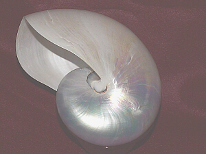
Whole Nautilus Shell
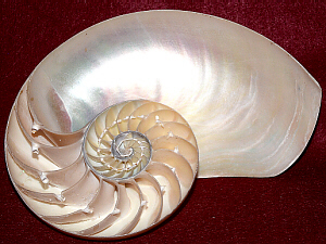
Nautilus Cut Open to View Chambers
Holes Between Chambers
While squid and octopus lack an external shell, the nautilus has a shell
thats divided into chambers, and the animal lives only in the most recent
chamber. The nautilus shells in these photos have had the outermost,
brown-and-white patterned layer
of their shells removed to reveal the pearlescent layer underneath. Notice
the row of holes between the chambers. These, or more correctly, the tube
that would be passing through them, is/are called siphuncles
and are involved in regulating bouyancy by changing the amount of water vs.
air present in the sealed chambers of the shell.
The squid has a region with arms and
eyes thats called its head. If a
fresh or preserved squid is available, note the number of arms and note the
suckers on them. The two arms that are longer help to capture prey, are
retractile, and may be called tentacles. In the center of the circle of
arms is the mouth with a beak thats shaped like that of parrot.
On the head, above the tentacles are a pair of well-developed
eyes. Adjacent to the head, on one side, is a muscular tube, the
funnel, with an opening at its tip (remember, the head and funnel
make up the foot of the squid, thus, technically, are ventral with the head
anterior and the funnel posterior).
The rest, the major portion, of the body is a dorsal hump, the
mantle, which encloses the internal visceral mass. It is extended laterally
to form a pair of triangular fins. Near the head, the mantle forms a loose
collar around the visceral mass. The gills lie in the mantle cavity.
Water from the mantle can be forced out the funnel for
propulsion. Also, a sac in the mantle cavity contains an inky substance,
which can be expelled in the jet as the squid or octopus is fleeing from
a predator.
Cephalopods have a well-developed nervous system, especially
a large brain with several pairs of ganglia between the eyes. They have
excellent vision, and octopuses are extremely intelligent and good at
problem-solving as in, seeing the feeder fish in the aquarium next door,
lifting the lid to squeeze through the filter and get out of the home
aquarium, using ones suckers to walk across the wall to travel to the
neighboring aquarium, getting into that neighboring
aquarium, eating all the fish, then going back home, and remembering to
close the lid so those pesky humans cant tell who ate the fish.
Octopuses have demonstrated tool use.
Octopus blood contains hemocyanin, the center of which
is a porphyrin ring with a copper atom in the center. This gives the blood
a blue color.
If available for
examination, compare squid and octopus how are they similar, and how are
they different?
Phylum Echinodermata
(echino = hedgehog, sea urchin, spiny; derm =
skin)
This phylum includes
- Class Asteroidea (aster = star) which includes the
starfish and sea stars
- Class Ophiuroidea which includes the brittle stars
- Class Echinoidea which includes the sea urchins and sand
dollars
- Class Crinoidea which includes the sea lilies or crinoids
- Class Holothuroidea (holo = whole) which includes the
sea cucumbers
Members of this phylum share several traits in common with,
and thus, it is believed are evolutionarily more closely related to
the chordates (vertebrates) than to other invertebrate phyla. They have an
endoskeleton (internal skeleton endo = within, inner)
covered by a thin layer of epidermis (skin), vaguely reminiscent of our
endoskeleton with skin on the outside. Also, the larval forms in these two
phyla are similar to each
other (and different from other animals). It is believed that these
similarities are evidence of a close evolutionary relationship between these
two phyla, even though that may not be obvious by looking at them.
All echinoderms are marine. The adults possess radial
symmetry, but it is recognized that is secondary and is superimposed on the
bilateral symmetry of the larvae (thus, is not a primitive trait).
Their larvae have bilateral symmetry, but metamorphose to adults that have
secondarily radial symmetry (have lost bilateral symmetry, rather than never
had it).
An important characteristic of this phylum is that the members
possess an unique hydraulic system, including external, suction-cup-like
tube feet connected by internal plumbing. By pumping fluid from one
part of the hydraulic system to another, echinoderms can create a strong
suction in the tube feet, aiding in locomotion and feeding.
Examine any plastic mounts
and/or skeletons of echinoderms that are available.
Class Asteroidea

Young Starfish, w.m.

Starfish Ray, x.s.
The young starfish in the left picture is unusual among starfish in that it
has six arms, rather than five. Many of its tube feet and its
sieve plate are visible.
Especially noticeable in the photo on the right are some of the tube feet.
Members of this class are called starfish (aster =
star), having five arms radiating from a central disk. The oral
surface (the side bearing the mouth) is the bottom, and that side has
furrows extending from the center out into each of the arms. On the
aboral surface (top), between two of the arms on the central
disc, is a whitish sieve plate which opens into the water
vascular system, the hydraulic system that enables the tube feet to
function. In the center of the sieve plate is a tiny anal opening.
On/in the skin, between the spines, are small skin
gills and pincer-like structures that help keep the skin free of debris
(but these are both very small and hard to see).
On the oral surface, rows of tube feet (which are part
of the water vascular system) extend from the ambulacral grooves (the
furrows in which they originate). The tip of each tube foot is a suction
cup. There are moveable spines along the margin of the grooves. As
previously mentioned, the grooves converge toward the central area, and
there, the mouth is found, surrounded by a soft, circular membrane.
Starfish feed on bivalves, such as oysters and clams. They
use hydraulic pressure in their tube feet to pull open the shells of these
bivalves. The starfish fastens onto the shell of the bivalve with its tube
feet and pulls the shell open, and since this doesnt involve the use of any
(or at least extremely few) muscles, the
hydraulic system of the starfish doesnt get tired, and since they have so
many tube feet, those can take turns so the tiny muscles in them can rest.
In contrast, the bivalve must use the previously-mentioned anterior and
posterior adductor muscles to try to keep its shell closed, and muscles cant
work forever without getting fatigued, so eventually those muscles tire and
relax, and the starfish is able to open the shell. After the
starfish has succeeded in opening the shell, the starfishs stomach, a large,
membranous sac, is everted through the mouth to cover the fleshy part of the
bivalve. The stomach secretes digestive juices that break down the bivalve,
and the mostly-digested food is taken into the stomach for further digestion.
Since little indigestible food is taken into the stomach, the remainder of
the digestive tube and the anus dont have a lot to do, and thus, are are
very small.
The digestive organs, etc., lie within a coelom filled
with coelomic fluid. The nervous system is simple, consisting of
a ring of nerves with nerves radiating into each arm.
Starfish can also reproduce asexually by regeneration
from chopped off arms. Starfish prey on oysters (valued as a human food
source), and years ago
before their regenerative powers were realized, the oyster fishermen would
destroy any starfish they found by chopping them up, then
tossing them back into the water. The chopped up starfish arms would just
regenerate whole new starfish, and the problem with starfish eating the
(desirable-to-humans) oysters intensified.
Starfish are either male or female. They release large
quantities of eggs and sperm into the open water, where fertilization takes
place. Starfish larvae are bilaterally symmetrical and look nothing like
the adults. They are free-swimming. Eventually, each larva attaches itself
to a convenient substrate and metamorphoses into an adult starfish.
Other Classes
Other classes in this phylum include Class Ophiuroidea =
brittle stars, Class Echinoidea = sea urchins, Class Crinoidea = the sea lilies
and feather stars, and Class Holothuroidea = sea cucumbers. While live sea
lilies (AKA crinoids) are not very numerous or common, now, and are found
only deep in the oceans, they were abundant in the Paleozoic Era and are
commonly-found fossils.
Phylum Chordata
There are other subphyla besides Vertebrata within Phylum
Chordata! These include Subphylum Hemichordata = tongue worms, Subphylum
Tunicata = the tunicates and Subphylum Cephalochordata = the lancelets.
While many of the traits in these groups vary, there are several things,
common to all chordates, that they and members of Subphylum Vertebrata all
share. At least in some stage in their life cycles, all possess
a stiff axial rod called the notochord, hence the phylum name.
Additionally, all chordates possess, at least in some stage of their life,
a dorsal, hollow, nerve cord and pharyngeal slits.
The nerve cord is worth special mention here. Annelids and
arthropods have a nerve cord that is ventral, solid, and of mesodermal
origin. Thus the fact that the chordate nerve cord is dorsal, hollow, and
arises from an invagination of ectoderm tissue points to a considerably
different evolutionary history.
Subphylum Cephalochordata
Note that members of this subphylum are in Phylum Chordata,
but are not in Subphylum Vertebrata not all vertebrates are chordates! In
general, members of this subphylum are known as lancelets or lancets for
their resemblance,
shape-wise, to that medical tool. The best-known member of this subphylum
is Amphioxus (Branchiostoma lanceolatum; amphi = on both sides,
double; branchi = gill, fin, hoarse; stoma = mouth), which
lives in shallow marine waters, buried in the sandy bottom with only its
anterior end protruding.
Like other chordates, members of this subphylum possess, at
least in some stage of their life cycles, a) a stiff axial rod called the
notochord, b) a dorsal, hollow nerve cord that is formed by
invagination of ectoderm tissue, and c) pharyngeal slits.
Class Leptocardii

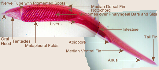
Amphioxus, w.m., Unlabeled and Labeled
Running down the sides of an Amphioxus are segmental, V-shaped
muscles called myotomes. The body is more flattened (side-to-side)
ventrally than dorsally. The head end is more flattened and somewhat less
pointed than the posterior end.
The membrane called the oral hood makes up the
anterior part of the head. Its bottom border is fringed with tentacles,
and the area enclosed by the hood and tentacles is called the buccal
cavity.
There is a median dorsal fin that runs all the way down
the back. Ventrally, from the oral hood to about two-thirds of the way to
the posterior end of the body run a pair of metapleural folds.
Ventrally, at about two-thirds of the distance, posteriorly, is the
atriopore, and the median ventral fin continues posteriorly
from there.
At the posterior end, the median dorsal and median ventral
fins expand to form the tail fin. The anus opens just anterior to the tail
fin, near the ventral fin.
Sometimes, in the anterior regions, ventrally, below or at
the bottom ends of the myotomes, whitish masses may be seen
these are the gonads.
Internally, the mouth is at the rear end of the buccal
cavity, surrounded by velar tentacles. From the mouth to about
half-way down the body are the pharynx and pharyngeal gill
slits. Behind the gill slits, the digestive tract narrows to form the
intestine, which extends back to the anus. In the region where the
pharynx intestine meet, extending anteriorly, is a dead-end tube that is the
liver. Above the digestive tract, extending end-to-end is a rod that
may appear yellowish on a prepared slide this is the notochord.
Just above that, also extending end-to-end is the dorsal nerve chord, which
is smaller in diameter than the notochord, and is hollow. It bears some
pigmented spots (near anterior end) which may be light-sensitive. Ventrally
and between the pharynx and body wall, and also behind pharynx is a body
chamber called the atrial chamber. This collects water that has
passed through the gill slits, and empties it to the the outside via the
atriopore.

Amphioxus, x.s. of Pharyngeal Region
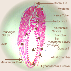
Amphioxus, x.s., Labeled
The view, above, is a cross-section through the pharyngeal area, and shows
many of the body parts mentioned, above. Throughout much of the pharyngeal
area, the atrial chamber is more expanded below and somewhat to the sides
of the pharyngeal area, itself, while this slide/photo is from an area where
the atrial chamber looks like a relatively thin area between the pharynx
and the outer layers of muscle and skin. Also, a cross section from another
portion of the pharyngeal area would show paired gonads to each side of the
pharynx, but in this view, there is only a small area on the right that
might be the end of one of the gonads. Notice how the pharyngeal
slits and branchial bars alternate around the pharyngeal cavity. The bars
are for support/structure, while the slits allow water to flow from the
pharyngeal cavity into the atrial chamber (and, eventually, out the
atriopore). For comparison, while we do not have pharyngeal slits as adults,
we do have them during a portion of our embryonic development.
Examine the plastic mount
of a whole Amphioxus. Examine the prepared slide of a whole mount of
Amphioxus (Carolina #Z2706) and of a cross section through the pharyngeal
region (Carolina #Z2720). Draw and label all parts that are visible.
Amphioxus has its own song! A number of recordings of this
song are available online. For example, see
Subphylum Vertebrata
Taxonomically, Subphylum Vertebrata would fit here. More
information on them can be found on the
cat Web page.
Other Things to Include in Your Notebook
Make sure you have all of the following in your lab notebook:
- all handout pages (in separate protocol book)
- all notes you take as you read through the Web page and/or
during the introductory mini-lecture
- all notes and data you gather as you perform the lab
- labeled drawing(s) (yours!) of any/all organisms
examined
- all plastic mounts
- all microscope slides
- all live, dead, and/or preserved specimens
- answers to all discussion questions, a summary/conclusion in your
own words, and any suggestions you may have
- any returned, graded pop quiz
Copyright © 2012 by J. Stein Carter. All rights reserved.
Based on printed protocol, background, and additional
information Copyright © 2012 J. L. Stein Carter
Chickadee photograph Copyright © by David B. Fankhauser
This page has been accessed  times since 25 Dec 2012.