Vertebrate Anatomy
Phylum Chordata, Subphylum Vertebrata:
In this lab, you will explore the internal anatomy of the cat,
thereby gaining a better understanding of the anatomy of other vertebrates,
including our own internal anatomy.
The internal anatomy of the cat is representative of the
anatomy of other vertebrates and is especially similar to that of the human,
more similar than other commonly dissected animals such as fetal pigs. These
cats belong to our Anatomy and Physiology students and we will be just borrowing
them to look at them. Thus, when you are finished, return the organs to
their correct positions, close the peritoneal cavity, and wrap the skin
closely around the cat. Slide the cat, head first, belly down into the
plastic bag. Note that these cats are triple-injected the arteries
have red latex in them, the veins have blue, and the lymphatic system has
yellow. Please use only a blunt probe and/or your fingers to move organs
around so you dont poke or tear anything.
Characteristics of Phylum Chordata include:
- a notochord (noto = the back; chord = string), which is
present in at least all embryonic/juvenile stages (may not be present in
adults of some subphyla/classes)
- a dorsal, hollow nerve cord formed by an infolding of ectoderm tissue
(compare with Annelida and Arthropoda which have ventral, solid, mesodermal
nerve cords)
- pharangeal slits (pharynx = throat), which originally functiond in
filter feeding, but in fish became modified as gills, and in mammals as our
eustachian tubes and ears
- a postanal tail (post = behind, after; anal refers to the
anus), which extends beyond the anus in many taxa (thus the anus isnt at
the posterior tip of the body) in humans, the tail is present during
embryonic development but is subsequently resorbed
Characteristics of Subphylum Vertebrata include:
- an endoskeleton comprised of bones and which is able to grow along with
the animal bones are calcified in many, but not all, classes (sharks bones
are not calcified)
- specific bones called vertebrae, which surround the nerve cord
- a definite head
- a brain enclosed within a skull
- a closed circulatory system with a ventral heart (number of chambers within
the heart varies
- excretion via kidneys
- separate males and females with sexual reproduction in most (a few groups
exhibit parthenogenesis and/or the ability to change sex midway through
the organisms lifetime)
More information about Phylum Chordata can be found at the
Phylum Chordata page.
Safety Considerations:
Due to the toxic chemicals (of course theyre toxic they
kill bacteria, dont they?) used to preserve these cats, these safety
precautions need to be observed:
There was a problem in the past with an unexpected squirt
of preservative liquid landing in the eye of a student who was not wearing
goggles. Trust me, you do not want to have to go through the ordeal of
being subjected to the eyewash station (and hospital emergency room) just
for that. WEAR GOGGLES when examining the cats. If you do need to
temporarily remove the goggles to draw/write in your lab notebook, do not
touch, move, prod, or get too close to the cat until you first replace the
goggles!
You should wear exam gloves (if you have a latex allergy, we
do have nitrile gloves, and a few vinyl gloves for people who are also
allergic to nitrile, available) when handling the cats, and
thoroughly wash your hands after removing the gloves. It is suggested that
you keep your gloves on while you put the cat away and wash the table and any
equipment used. When everything else is done, then remove your gloves and
wash your hands. Obviously, if a glove gets a hole in it, it should be
replaced, but please do not waste gloves by constantly removing them just
to draw a picture or something, then tossing them and getting a new pair. If
you do need to temporarily remove a glove, if at all possible, please
carefully remove it so that you can put it back on. Please, as much as
possible, attempt to limit your glove consumption to one pair per lab
period.
Cat parts should not be disposed of in the regular trash.
Since we are borrowing cats from the A&P students, all cat parts should
be returned to the bags with the cats, but in the unlikely event something
should need to be thrown away, there is a special red, biohazard bag in a
special red, biohazard trash can, into which that should be placed.
While there is no prohibition against doing so, there is no
need to disinfect the tabletops after working on the cats!
The point of all the preservatives in the cats is to be so toxic that they
totally prevent the growth of any bacteria. Thus, any liquid left on the
table after working on a cat is not cat juice, but rather is excess of
those (toxic) preservatives. However... you do need to
thoroughly wash the tables with warm, soapy water to remove all the
preservative as well as any cat crumbs and fur, and then rinse the tables
and the sponge you used to remove all the soapy water. DO NOT LEAVE FUR
AND CRUMBS OF TISSUE IN THE SINK! (Collect them and put them in the
red, biohazard bag.) If you have sponged most of the water off the tabletop,
it should dry fairly-quickly on its own without the need to use a paper towel,
but if, for some reason, you do really need to use a paper towel to dry it,
please DO NOT get a whole handful of paper towels. Rather, just use one (or
two?).
Any dissecting tools (blunt probe) used should be washed with
warm, soapy water, rinsed, thoroughly dried, and then returned to their
proper storage location.
Pregnant women are encouraged to read the MSDS for the
preservative used on the cats in order to decide what safety precautions
they wish to take. That probably should include not working directly over
the cat at close range where one would be likely to breath a lot of
preservative fumes.
Materials Needed:
- goggles and exam gloves
- previously-dissected cats from the A&P class
- blunt probe
- possibly, optional books (in addition to your protocol book and this Web
page?), to help identify internal organs
External Anatomy:
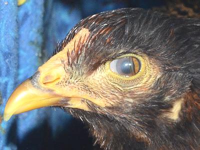
Stormys Inner Eyelid
Many animals have a third eyelid which sweeps across the
eye to clean it, or in some animals, can be closed to protect the eye in
a dusty situation. In humans, all we have left of this is the pink lump
in the inner corner of our eye.
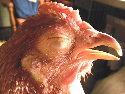
Frodo Being Sleepy
As you know, when we humans close our eyes, it is our upper
eyelid that goes down. Interestingly, in chickens, their lower eyelid goes
up.
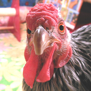
Sally Looking Forward
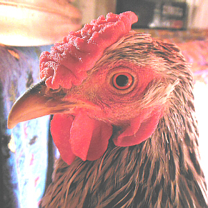
Sally Looking Sideways
An animal that is potential prey for another animal has its
eyes on the sides of its head and the eyes operate independently, giving the
animal nearly 360° vision to better watch for danger. A
predator
has its eyes on the front of its face, giving it excellent
binocular
vision for depth perception and judging distance to prey. An interesting
combination of these traits can be found in a chameleon (not an anole).
Chameleons eat insects, so need binocular vision to capture dinner, but are
also potentially dinner for someone else. They have their eyes on the sides
of their heads, but the eyes stick out and can swivel around. Chameleons can
use their eyes independently to watch for predators, yet when a potential
meal hops into sight, can focus both eyes on the insect to judge the
distance before flicking out a sticky tongue to catch it. Interestingly,
because of the location and mobility of a chameleons eyes, it can
rotate its eyes backwards, and have binocular vision behind its head!
Chickens, also, have their eyes on the sides of their heads, and they work
independently to watch for predators, but chickens use their binocular
vision to focus on the food theyre about to pick up.
Internal Anatomy:
Examine the preserved and dissected cat, and locate the
following features. Review with your lab partner the function of each
feature as you locate it. In your lab notebook, make labeled
illustrations of 1) neck and thorax, 2) abdomen, and 3) ventral view of the
brain, indicating the location of the features listed below. Also, take
notes on locations, functions, etc. of the various organs/systems that
you examine. Take the time to think about how what youre seeing compares
with what you already know about your own body, and write down your thoughts.
Note that while,
here, the organs are sorted/discussed by system, your drawings should be of
the body regions just mentioned, and should include all organs/systems
located within that body region.
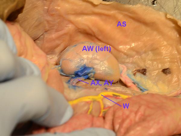
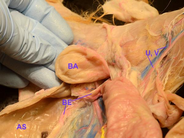
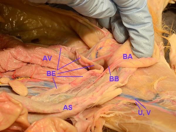
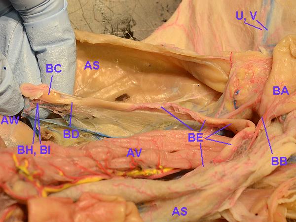
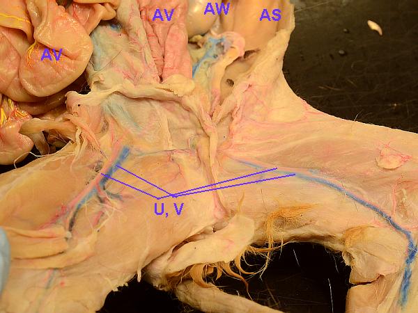
The letters in the list of body parts correspond to the labels on the photos,
included below the list.
More information on tissues and body system can be found on the
Tissues, Organs, and Systems page.
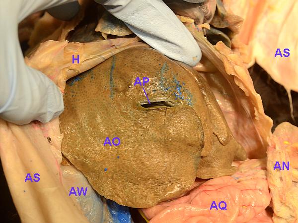
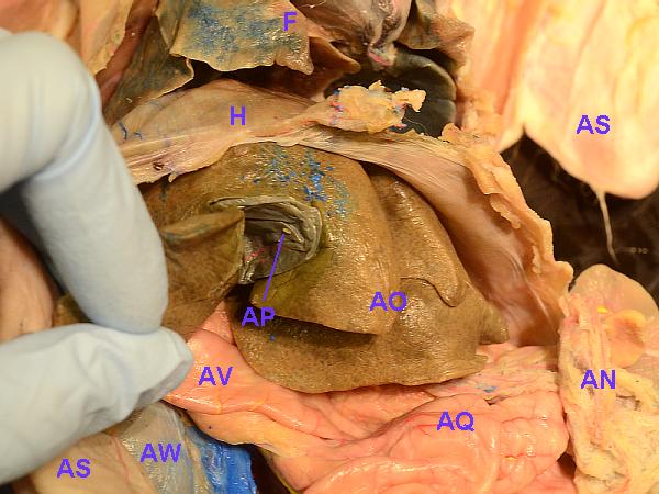
- Respiratory System
- nasal
cavity: This warms, moistens, and filters the air. The sinuses
are convoluted, narrow passageways.
- nasal
and oral pharynx: (pharynx = throat). Note proper
pronunciation:
fair-inks.
- larynx:
This is also known as the voice box. The epiglottis (epi = upon,
over) covers the larynx.
Note proper pronunciation:
lair-inks.
The cricoid cartilage is between the larynx and
trachea.
- trachea:
(trachea = windpipe) This has cartilaginous rings (similar to
those in a shop-vac hose) to hold it open.
(The esophagus is the muscular tube right behind and parallel to
it.)
- bronchi:
The bronchi (singular = bronchus) are the two main branches from the trachea to the lungs.
These are located behind the heart. An inflammation of the bronchi is
called bronchitis.
(bronch = windpipe)
- lungs:
Cats lungs have 3 lobes on each side, whereas humans lungs have 2
lobes on the left side and 3 on the right. This may relate to the fact
that, while our heart is located in the center of our chest, it does
lean a bit toward the left.
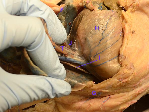
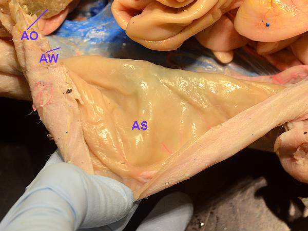
- pleura:
The thoracic cavity is lined by pleural membranes (pleura =
rib, the side), which is smooth and slippery. Parietal pleura
lines the outside wall of the chest, while the visceral pleura coats
the organs (pariet = a wall; viscer = the organs of the
body cavity). An inflammation of the pleura is called pleurisy: the
inflamed membranes dont slide well, but rub against each other, so
it hurts to breathe.
- diaphragm:
In mammals (including cats and humans), this muscle layer
divides the abdominal and thoracic cavities (dia = across,
through; phragm = fence) and is used to pull air into
the lungs.
 However, that is not true of all other vertebrates. For example, a
frog opens its nostrils and expands the floor of its mouth to draw air
into its mouth. Then it closes its nostrils and uses the floor of its
mouth to push air into its lungs.
However, that is not true of all other vertebrates. For example, a
frog opens its nostrils and expands the floor of its mouth to draw air
into its mouth. Then it closes its nostrils and uses the floor of its
mouth to push air into its lungs.
Birds also do not have a diaphragm, so their liver and heart are
almost touching. If their liver becomes enlarged for some reason, it
can rub on the birds heart, a sound which a trained veterinarian can
hear. Birds have a number of air sacs which branch off their lungs.
Their chest muscles move their sternum (breast bone) outward,
thereby pulling air into the lungs and air sacs, and thus, it
is critically important that a pet bird not be restrained so tightly
that its chest cavity is unable to expand. Unlike us, they exhale by
contracting other muscles to put pressure on the air sacs to push
the air out.
The lungs, themselves, do not expand and contract like mammalian lungs.
Because of these differences, it takes two breath cycles for air to
move in, throughout, and back out of the respiratory system. (This
complex process is explained in greater detail on a number of other
peoples Web sites: do a Google search for how do birds breathe.)
- phrenic
nerves: This is a pair of nerves that regulate the diaphragm (one
on each side). These are white strings, the bottom ends of which
attach to the diaphragm. They run from the diaphragm,
along either side of the heart, to the neck, then come out where the
neck meets skull. (phreni = diaphragm)
More information on the respiratory system can be found on the
Respiratory System page.
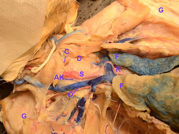
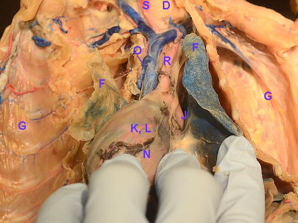
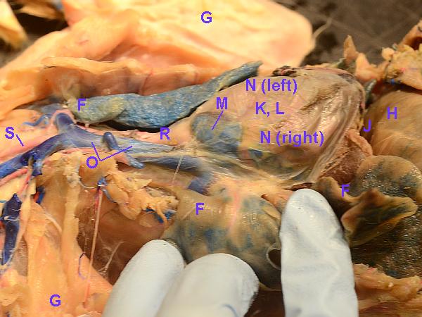
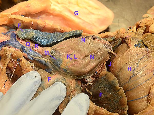
- Cardiovascular (Circulatory) System
- mediastinum:
This is the area between lungs: it contains the heart and thymus
- heart:
Fish have a two-chambered heart (one atrium and one ventricle) which
pumps blood to both the body and the respiratory system (gills).
Amphibians and most reptiles have a three-chambered heart (two atria
and one ventricle), so their heart separately receives blood from the
body and
from the respiratory system, but the blood mixes in the ventricle
and is pumped back out to both the body and the respiratory system.
Crocodiles (which are reptiles), birds, and mammals have
four-chambered hearts (two atria
and two ventricles). However, the evolutionary history of the
crocodilian heart differs from that of the avian and mammalian hearts.
In a four-chambered heart, the right atrium receives blood from the
body. That is sent to the right ventricle, which pumps it to the lungs.
The blood is returned from the lungs to the left atrium, and from
there it goes to the left ventricle. The muscular left ventricle
sends the blood out to the body.
- pericardium:
This is the membrane around the heart. It is slippery and lubricated
so that the heart can move freely as it beats.
(peri = around, cardio = heart)
- atria:
(singular = atrium) Mammals have two: a right and left atrium or
auricle (atrium = vestibule, entrance hall; auricle =
little ear). These are the upper portions of the heart and receive
blood back from either the body (right) or lungs (left).
- ventricles:
These are the lower, muscular portions of the heart and receive blood
from the corresponding atria. Birds and mammals have two ventricles,
and the left one is especially muscular to send blood to whole body.
- superior
vena cava: This delivers blood from the head and arms to the right
atrium.
- inferior
vena cava: This delivers blood from the lower body to the right
atrium.
- pulmonary
trunk: The pulmonary trunk (or pulmonary artery) carries
deoxygenated
blood to the pulmonary arteries (which go to the lungs). It comes
from the right ventricle, out on top of heart, then divides and goes
to the lungs. The pulmonary vein carries oxygenated
blood back to the heart from the lungs. (Note: arteries/veins are
defined/determined by the direction of blood flow [arteries rarr; away],
not by O2-content. The umbilical artery also
carries deoxygenated blood away from the baby to the placenta, and
the umbilical vein carries oxygenated blood from the placenta back
into the babys body.)
- aorta:
This looks yellowish because it is so muscular/elastic. The first,
big portion that goes up and over is the aortic arch.
(aorta = heaving)
- carotid
arteries: These are among the first to branch off the aorta.
They go up to the neck, pass by the larynx, and go up into the head.
These are large, red (because filled with latex), and carry the
majority of blood to the brain.
- descending
aorta: This is the portion of the aorta which goes down behind the
left lung, through the diaphragm, and into the abdomen.
- common
iliac arteries (left and right): These branch off the descending
aorta where it ends at the hip (ilium = the groin, flanks).
These are red because they are filled with latex. Each branches
into a group of femoral (deep, superficial, common) arteries.
- common
iliac veins: These run parallel to the iliac arteries and are
filled with blue latex. They join to form the bottom end of the
inferior vena cava (which goes to the heart).
More information on the circulatory system can be found on the
Circulatory System page.
- Lymphatic System and Immune System
- lymphatic
system: This contains yellow latex. Its job is to filter fluid
to remove bacteria, etc. Especially noticeable to the outside of the
digestive and circulatory systems is a big, yellow trunk up to the
left subclavian vein.
- lymph
nodes: These yellow (laytex-filled) nodes may be easily found in
the mesenteries that support the intestines. Other, less-obvious
lymph nodes are found throughout our bodies. Because of their
filtering functions, along with the bacteria that they trap and
remove, they are also noted for their ability to trap metastasizing
cancer cells, which then may start to grow in the lymph node. Thus,
a lymph-node biopsy is often performed to determine how far someones
cancer might have spread.
- thymus:
The thymus, which is located in the thorax, activates T-cells to kill
foreign tissue. It is especially large and obvious in childhood, but
smaller in adults.
- spleen:
This is located on the left side of the abdomen, by the stomach. It is
involved in recycling blood cells, and is famous for becoming enlarged if
someone has mononucleosis.
More information on the immune system can be found on the
Immune System page.
- Endocrine System
- thyroid
glands: These are small and located, up near the larynx. Humans
have one, united one, but
in the cat, they are separate and are located on either side of
larynx. The hormones they make regulate metabolic rate. They absorb
iodine and make thyroxine.
- adrenal
glands: In the cat, they do not sit directly on the kidneys, but are more
medial (closer to the center) and are yellowish. Adrenal glands are
the source of adrenalin (ad = to, toward; renal =
kidney) and a number of other hormones.
Note that the pancreas, testes, and ovaries, all described below, also serve
endocrine functions.
More information on the endocrine system can be found on the
Endocrine System page.
- Nervous System
- brain:
This is surrounded by three membranes called meninges: the outer one is the
dura mater (dura = hard; mater = mother). Below, the
major parts of the brain are described.
- cerebrum:
Most of the brain is comprised of these two hemispheres (hemi =
half). These control thought, motion, speech, etc., and each
hemisphere controls the opposite half of the body.
- cerebellum:
This is located behind the cerebrum and is the center of skilled physical
movement and pattern. Someone who has had a stroke in his/her cerebellum
may be able to move, but not put a series of movements together in a
meaningful, useful way: for example, may not be able to pick up a fork,
then use that to pick up some food and transfer it to his/her mouth.
(cerebell = brain; -um = structure, tissue, thing)
- medulla
oblongata: This is the center of involuntary, life-sustaining functions
such as breathing, heart rate, and vomiting. It is the inferior-most
portion of the brain. (medull = marrow, pith)
- pons:
This is a bridge helps to connect the right and left sides of the
cerebellum. (pons = bridge)
- optic
nerve: You will probably observe this as cut-off stumps which form an X"
under the brain. The optic nerves cross, here, to the other side of the
brain.
- olfactory
tract: This anterior-most portion of the brain is involved in our sense
of smell, and can link those smells to past events such that a given smell may
instantly and vividly evoke recollection of memories.
Note that the phrenic nerves, described above, also are
part of the nervous system.
In general, the larger an animal is, the larger and more complex its brain
often is. However, there has been some interesting research done on birds
brains, which while considerably smaller than those of an equivalent-sized
mammal, are wired just differently-enough that they are more efficient,
often making a bird smarter than an equivalently-sized mammal.
More information on the nervous system can be found on the
Nervous System page.
- Digestive System (and associated abdominal organs)
- salivary
glands: There are several of these, located in the sides of the
face near the jaw, and connected by ducts beside the tongue.
Salivary amylase is the main enzyme in saliva.
- esophagus:
The esophagus runs posterior to the trachea and penetrates the diaphragm
to connect to the stomach.
- stomach:
The stomach starts at (is attached to) the diaphragm, and is a J-shaped
bag. The main enzyme it produces is pepsin, and its average pH is
about 1 to 1.5.
- pyloric
sphincter: This is a constriction/valve at the bottom of the stomach,
between the stomach and the duodenum. (pylorus = a
gate-keeper)
- greater
omentum: Not a functional part of the digestive system, per se,
but connected to it, this is a fat-filled skin which is attached to
the stomach, and from there, lies ventrally over the intestines and
other abdominal organs. It helps to insulate and protect the abdominal
contents. (oment = fatty skin)
- liver:
This is the major metabolic organ in the body. It makes bile for fat
digestion (bile emulsifies fat), and that bile is stored in the gall
bladder prior to being secreted into the small intestine.
- gall
bladder: This is the dark (raisin-like) spot in the center of the liver
(the human gall bladder is below/behind the liver). Its bile duct connects
it to the duodenum.
- duodenum:
This is the first curve of the small intestine. The inner surface is
covered with villi (fingers) to increase its surface area for better
absorption and which give it a velvety appearance. (villi =
shaggy hair)
- pancreas:
This is grayish-brownish, diffuse and not really obvious if you dont
know where to look for it. It is located in the folds of the
mesenteries, near the duodenum. It secretes digestive enzymes into the
duodenum. It also serves an endocrine function, secreting glucagon
and insulin to regulate blood-sugar level.
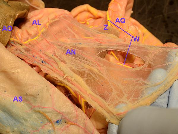
- peritoneum:
This is similar to the pleura mentioned above, with the difference that
it is located in the abdomen (while pleura is located in the thorax).
Visceral peritoneum covers the intestines, while parietal peritoneum
lines the outer wall of the abdominal cavity.
- mesenteries:
The mesenteries are all the transparent membranes connecting and
holding in place the intestines. The pancreas and a number of lymph
nodes are located within the mesenteries.
- ileocecal
valve: This is the junction between the last part of small
intestine and the colon. (ileum = twisted)
- colon:
Cats have a colon and cecum, but cats do not have an appendix. The
colon is also known as the large intestine, and is shaped like a
question mark (?). The cecum is a dead-end region by the
junction with the small intestine (cec = blind), and in humans,
the appendix is attached to the blind end of the cecum. The rectum
(rect = straight) is the end of colon where feces are stored
until eliminated.
More information on the digestive system can be found on the
Digestive System page.
- Excretory (Urinary) System
- kidneys:
Osmosis occurs in the kidneys. They are connected by the ureters to
the bladder.
- renal
arteries: These supply the kidneys with blood, bringing them
both nourishment and excess water and nitrogenous wastes to filter.
- renal
veins: The diameter of the renal veins is bigger than that of the
renal arteries because the veins carry blood under lower pressure.
- ureters:
These carry urine from the kidneys to the bladder.
- urinary
bladder: This stores urine until it is voided.
- urethra:
This carries urine from the bladder to the outside. In males, the vas
deferens connects with and empties into the urethra, so that both
semen and urine can be voided via the penis.
More information on the excretory system can be found on the
Excretory System page.
- Reproductive System
- ovaries:
These are present in females and produce eggs.
- fallopian
tubes: These are present in females and transport eggs to the
uterus. In birds, the fallopian tube has the extra functions of adding
the egg white and shell around the egg/embryo. Note that, as in
humans, to turn into a chick, the egg must first be fertilized by
sperm which have traveled up to the vacinity of the ovary.
- uterus:
This is present in females and is where embryos develop
into babies. In humans, most females have one, central uterus
in which (in most cases) one baby at a time develops. In cats, the
uterus is V-shaped, allowing for numerous embryos to simultaneously
attach at different points, resulting in a litter of kittens born
at the same time. In birds, reptiles, etc., the zygote/embryo (AKA
fertilized egg) is packaged into an egg shell, released from
the females body, and develops externally. We placental mammals
whose young develop within the mothers uterus are the exception
rather than the rule.
- vas
deferens: This is present in males and joins the urethra to pass
semen to the penis (in animals whose males have that organ birds,
for example, do not).
- testes:
These are present in males and make sperm. They develop in the
abdomen, in about the same location as ovaries do in females, then
during embryonic deveopment, move
downward and pass through the inguinal canal (just under the skin) and
out into the scrotum. This is important because a) undescended testes
are more prone to developing cancer and b) the sperm they produce need
a slightly cooler environment than internal body temperature.
- testicular/ovarian
arteries: These are the blood supply for the testes or ovaries.
The testicular or ovarian arteries originate above/near kidneys (during
embryonic development). In males, as the tested descend, their
arteries grow longer, grow down with the testes.
- testicular/ovarian
veins: These return blood from the testes or ovaries. Again, in
males, thes elongate and grow down with the testes.
Birds and some other vertebrates have a cloaca. This is an organ, a
space, into which the colon, the urethra, and the oviduct(s) or vas deferens
all open, and from which they all share a common opening to the outside.
More information on the reproductive system can be found on the
Reproductive System page and
the Reproductive Physiology page.
- Skeletal System
Key:
S = shoulder,
E = elbow,
W = wrist,
C = carpals (hand),
D = digits (fingers),
H = hip,
K = knee,
A = ankle (heel),
F = foot,
T = toes
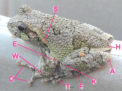
Amphibian (treefrog)
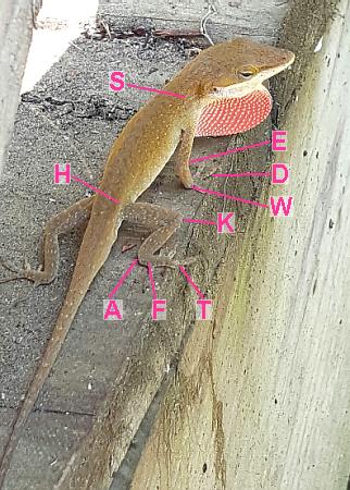
Reptile (Green Anole) © E. A. Stein
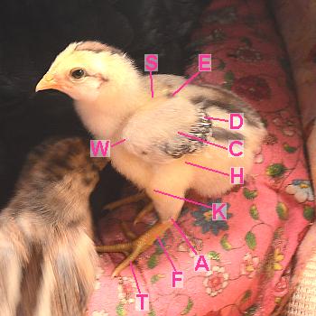
Bird (Chicken)
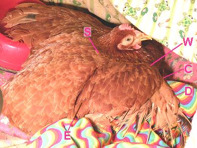
Bird (Chicken)
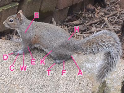
Mammal (squirrel)
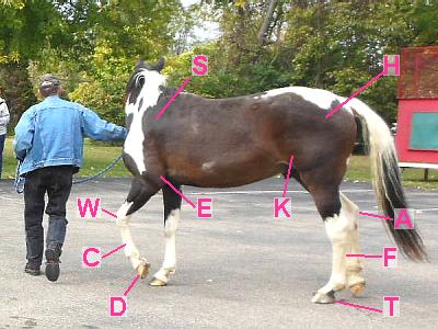
Mammal (Horse)
- skeleton:
Due to the way these cats have been dissected, you probably wont be
able to see any actual bones (maybe some ribs?). One important point
to make, here, is
that the skeletal structure of amphibians, reptiles, birds, and mammals
is pretty-much the same. They all have the same bones in the same places,
facing the same direction, as we humans do. Many people make the
mistake of looking at the heel of a dog or goat or chicken,
and think it is a backwards knee: it is NOT its a heel! They are
walking on tip-toe, their feet are a lot longer than ours, and their
tibia (shin bone) is shorter. Their
wrists, elbows, heels, knees, etc. are in the same positions and
facing the same directions as ours.
More information on the skeletal system can be found on the
Skeletal System page.
- Muscular System
More information on the muscular system can be found on the
Muscular System page.
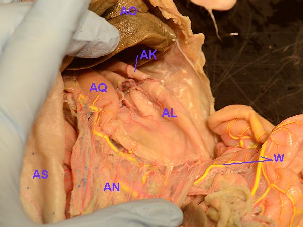
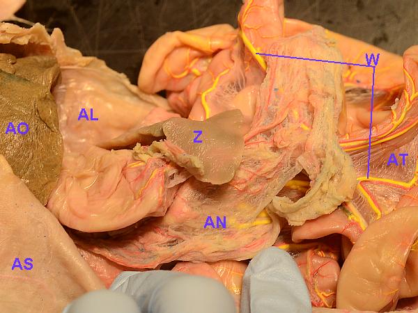
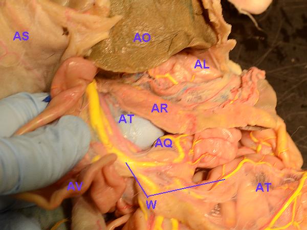
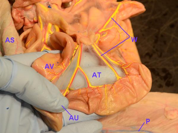
| Cat Internal Anatomy Thorax |
| Labels correspond to those of parts listed, above. |
 |
 |
 |
 |
 |
 |
| Cat Internal Anatomy Abdomen |
 |
 |
 |
 |
 |
 |
 |
 |
 |
 |
 |
 |
 |
 |
Clean-Up:
IMPORTANT: When you are finished, return the organs
to their correct positions, close the peritoneal cavity, and wrap the skin
closely around the cat. Slide the cat, head first and belly down, into its
plastic bag. Press the air out, fold the bag over lengthwise, and secure the
end with three turns of a sturdy rubber band. Return the bagged cat to the
storage bag in the box. All cats should be headed the same direction in the
box (all rubberbands on the same end of the box).
Clean up stray pieces and fur. As mentioned above, wash the
desk top with soap and water, then rinse. Make sure any stray fur or
cat crumbs are placed into the red bag. MAKE SURE TO CHECK THE DRAIN IN
THE SINK YOU USED!
Other Things to Include in Your Notebook
Make sure you have all of the following in your lab notebook:
- all handout pages (in separate protocol book)
- all notes you take as you read through the Web page and/or
during the introductory mini-lecture
- all notes and data you gather as you perform the lab
- labeled drawings (yours!) of
- ventral view of neck and thorax area
- ventral view of abdominal area
- ventral (and side) view(s) of brain
- answers to all discussion questions, a summary/conclusion in your
own words, and any suggestions you may have
- any returned, graded pop quiz
Protocol Copyright © 1987 D. B. Fankhauser
Background and additional information Copyright © 1995, 2017 by J. Stein Carter. All rights reserved.
Lizard (Green Anole) photograph Copyright © 2016 E. A. Stein
Chickadee photograph Copyright © by David B. Fankhauser
This page has been accessed  times since 18 Dec 2010.