 any
people who came to play volleyball brought their families.
any
people who came to play volleyball brought their families.
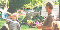 It was interesting to look at all the people and try to figure out, by looks,
which children were siblings or which adults were their parents. In some
cases, the genetic similarities were easier to see, while in other cases
the mixture of genes from the two parents made it more difficult to visually
see relationships between family members.
It was interesting to look at all the people and try to figure out, by looks,
which children were siblings or which adults were their parents. In some
cases, the genetic similarities were easier to see, while in other cases
the mixture of genes from the two parents made it more difficult to visually
see relationships between family members.
Mendelian and Human Genetics
There used to be a TV show whose punch line was always,
Smile! Youre on Candid Camera.

Can you tell which of these people are related? (Hint, look at
noses and cheeks as well as smiles.)
 Gregor Mendel, an Austrian monk, is considered to be the
Father of Modern
Genetics for the discoveries he made by breeding pea plants. Mendel figured
out that we get a copy of each gene from each parent. He also found that
some alleles (alternate forms for a gene) are dominant
while others are recessive, such that, if a person (or other organism) has
a dominant allele from one parent and a recessive allele for the same gene
from the other parent, the dominant trait will be expressed.
Gregor Mendel, an Austrian monk, is considered to be the
Father of Modern
Genetics for the discoveries he made by breeding pea plants. Mendel figured
out that we get a copy of each gene from each parent. He also found that
some alleles (alternate forms for a gene) are dominant
while others are recessive, such that, if a person (or other organism) has
a dominant allele from one parent and a recessive allele for the same gene
from the other parent, the dominant trait will be expressed.
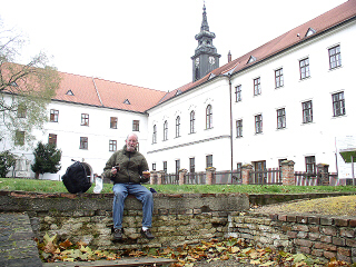
Here, Dr. Fankhauser is eating a picnic breakfast while sitting on the
remains of Gregor Mendels greenhouse in Brno, Czech Republic. A statue of
Mendel is to his left, between the bushes, back by the
monastery wall.
An individual who has two identical alleles (two dominant OR
two recessive
alleles) is called a homozygote, while someone who has two different
alleles (one of each) is called a heterozygote. Gametes (eggs and
sperm) have only one copy of each gene, therefore only one allele for
each gene, and when a sperm fertilizes an egg, the condition of having two
is restored. An organisms genotype is its actual genetic makeup
for the gene(s) under consideration (for example, heterozygous dominant
or homozygous recessive, etc.), while its phenotype is how those
genes are expressed (sort-of, What does it look like? except that not all
genes code for things that can be seen,
visually).

 To make it easier to keep track of genetic crosses, rather than having to
explain something like, I crossed a homozygous purple-flowered pea plant
with a homozygous white-flowered pea plant, and all the offspring were
heterozygous and had purple flowers,
geneticists came up with the idea of using letters to represent the various
alleles. Dominant alleles are represented by capital letters and recessive
alleles are represented by the same lower case letters. Thus, in our example,
if purple flowers are dominant and white is the recessive allele of that same
gene, we could use capital P (for purple) to represent purple and
lower case p to represent white. We could then rephrase our description
as, I crossed a PP pea plant with a pp plant and got Pp
offspring.
To make it easier to keep track of genetic crosses, rather than having to
explain something like, I crossed a homozygous purple-flowered pea plant
with a homozygous white-flowered pea plant, and all the offspring were
heterozygous and had purple flowers,
geneticists came up with the idea of using letters to represent the various
alleles. Dominant alleles are represented by capital letters and recessive
alleles are represented by the same lower case letters. Thus, in our example,
if purple flowers are dominant and white is the recessive allele of that same
gene, we could use capital P (for purple) to represent purple and
lower case p to represent white. We could then rephrase our description
as, I crossed a PP pea plant with a pp plant and got Pp
offspring.
Geneticists often use Punnett squares (named after
Reginald Crandall
Punnett) to keep track of genetic crosses. For example, suppose a heterozygous
brown-eyed man (brown is dominant) marries a blue-eyed woman, and we want to
figure out what eye color their children might have. If the man is heterozygous,
we could express his genotype as Bb (using B for brown), and the
womans genotype would have to be bb if she has blue eyes. When the
mans body is making sperm, half of his sperm will get his B allele
and half of his sperm will get his b allele. When the womans body is
making eggs, half of her eggs will get her b allele, and half will get
the other b allele. In a Punnett square, the possible types of sperm
are listed across the top and the possible types of eggs are listed down the
left side. A Punnett square is as big in either direction as the number of
possible gametes that parent can produce. In this case, since were dealing
with two types of sperm from the father, our Punnett square will have two
columns, and since were (sort-of) dealing with two types of eggs from the
mother, it will have two rows (yes, technically, we could get by with one row).
Thus, the initial set-up of our Punnett square would look like
this:
The boxes in the center represent what the possible childrens
genotypes would be if that sperm fertilized that egg. Thus, the genotypes of
the sperm are copied down the appropriate column, like
this:
Similarly, the genotypes of the eggs are copied across the
appropriate row, like this:
When its all put together, and both the sperm and
the eggs have been added, the final result looks like
this:
We can then see that half of their children would be
expected to be genotype
Bb or heterozygous dominant or heterozygous brown eyes, while the
other half would be bb or homozygous recessive or homozygous blue.
In terms of phenotype, we would say that half of their children should have brown
eyes and half should have blue. Understand, thats not saying everyone will
have exactly four children, but rather, its a prediction, an average. This
couple might only have three children, and perhaps two of those will have brown
eyes while the third has blue eyes. Maybe theyll have two children, both of
whom end up with blue eyes. Maybe theyll have four, three of whom will have
brown eyes and only one blue. (Its like, if you would toss a coin 100
times, you might not end up with exactly 50 heads and 50 tails, but your
results would probably be close to that.)
When you start to dig deeper, there are some interesting
inheritance patterns
that show up. As one example, humans and other mammals have a number of
regular chromosomes called autosomes plus a set of sex chromosomes.
Typically, females have two X chromosomes (genotype XX), while males have an
X and a Y (genotype XY), and just like all the other chromosomes, the X and
the Y have genes on them that code for various traits. However, the X chromosome
is much larger than the Y, and contains a number of genes that have no
counterpart on the Y chromosome. While the Y chromosome is much smaller than
the X, it actually does have a few genes on it that are not represented on the
X. Genes that are located on only the X or Y chromosome are
called sex-linked genes. For example, in humans, red-green colorblindness
and hemophilia are two traits that are carried on the X chromosome, while
one variety of six-fingeredness and hairy earlobes are carried on the Y
chromosome.
Sex is also a phenotype, and just like any other
phenotype, is a matter
of how the persons alleles are expressed. Its not merely a matter of
whether the person has two X chromosomes or an X and a Y, but rather, what
alleles the person has for the genes which are located on those chromosomes,
and how those alleles are expressed. An example, described in more detail
on the Biol. 105 Sex-Linked Genes Web page (link below), is the case of a genetic
condition called androgen insensitivity syndrome (AIS). On everyones
X chromosomes, there is a gene that codes for peoples cells to build receptor
sites to receive androgen (testosterone), and there is no corresponding gene
on the Y chromosome, so this is an X-linked or sex-linked gene. To briefly
summarize, a person with AIS has an X and a Y chromosome, but on that one and
only X chromosome, has an altered allele that does not allow the testosterone
receptor sites to form/function properly. Thus, even though that person, due
to the influence of other genes on the Y chromosome, has undescended testes
which make lots of testosterone, the rest of the persons body never receives
the testosterone message, and by default, other than the undescended testes
and a missing uterus, she develops totally, unambiguously
female traits. Often, girls with AIS dont even discover that they have it
until, despite otherwise normal development at puberty, they fail to menstruate.
The opposite is also possible: Ive been told there was a case where a couple was not
able to conceive a baby and thus were going through various testing to determine
why. The results of the tests showed that the very masculine, bearded husband
was, chromosomally, XX.
There are a number of other genetic and chromosomal
mutations which
can affect humans (see link, below). Among many others, these include such
things as
- Rh incompatability, which may occur when a Rh-negative mother is
pregnant with an Rh-positive baby,
- Sickle-cell anemia, in which abnormal hemoglobin causes red blood cells
to crinkle up (sickle),
- Down Syndrome, in which the person has an extra chromosome #21 (so there
are three in that pair,
- Huntingtons Disease, which is a progressive deterioration of the
nervous system,
- Cystic Fibrosis, in which there is a problem that causes large amounts
of mucus to be secreted in the lungs (and other areas of the body),
- Tay Sachs, which is a neurological disorder, and
- Achondroplasia, in which a heterozygous person is a dwarf and a homozygous
baby dies prenatally and is miscarried.
Background Information
Links to Related Information on Our Web Server
The following Web pages contain information related to
various aspects of genetics.
- Bio Lecture Genetics
- General information on Mendelian genetics and typical patterns of inheritance
- Bio Lecture Genetics Practice Problems
- A page of practice Punnett squares
- Bio Lecture Linked and Sex-Linked Genes
- Information on genes located on the same chromosome as each other and information on genes located on the X or Y chromosomes, including discussions of hemophilia and androgen insensitivity syndrome
- Bio Lecture Human Genetics
- Information on a variety of human genetic and chromosomal abnormalities
- Bio Lab Genetics Activity
- An activity using coin tosses to simulate genetic crosses
Your Assignment
Genetics Practice
There will be only one, combined assignment for this weeks
topics (mitosis and meiosis, genetics, and DNA). Thus even though this will
appear on each of those three pages to remind you, you only need to do it once.
Genetics is one of those things that just needs lots of practice to get it.
The grading criteria for this assignment are given below, and you should also
refer to those as you work on the assignment.
A total of 32 points is possible.
- Mitosis
- Read through the Biol. Lecture Web pages on mitosis and meiosis to
become familiar with those processes, how they are the same, and
how they differ.
- Find/collect a group of similar but distinguishable objects such as
coins, pieces of string or yarn, socks, or whatever is handy. These will
be used to represent the chromosomes in the nucleus of a cell. Also,
obtain several, longer pieces of string or yarn to represent cell and
nuclear membranes.
- Make a cell. Use a longish piece of string to make a circle to
represent the cell membrane. Use a shorter piece to make a smaller
circle inside to represent the nuclear envelope. This organism will
have 6 chromosomes (3 from the father and 3 from the mother). For
this you will need 3 pairs of something; for example a pair each of
black, red, and blue socks or a pair each of pennies, nickles, and dimes
(or whatever is handy that will suit the purpose). Put these 6
chromosomes into the nucleus of the cell.
- Just before mitosis happens, the chromosomes replicate, but the
halves (called sister chromatids) stay attached. Simulate this
by stacking 6 more identical
objects (well, come as close as you can...) on top of the existing
6 chromosomes. For example, stack another penny on top of each of
the two existing pennies, another nickle on top of each of the two
existing nickles, etc.
- In prophase of mitosis, one thing that happens is that the nuclear
envelope disintegrates. To demonstrate this, remove the string thats
the nuclear envelope and set it aside.
- In metaphase, all the chromosomes line up along the equator of
the cell. Line up your 6 chromosomes (each with its partner still
on top) in a row (single-file) across the middle (equator) of your
cell.
- In anaphase, the halves of the chromosomes separate and travel to
opposite poles of the cell. For each of your 6 chromosomes, now is
the time to separate the partners. For each of the 6 stacks of 2, move
one of the two items to the north pole of the cell and one to the
south pole of the cell. When youre done, each pole should have a
collection of 6 objects/chromosomes identical to the 6 with which you
began.
- In telophase, the nuclear envelopes re-form and the cell divides
into two. First, find a piece of string with which to form a circle
around each of the two groups of chromosomes to show the nuclear
envelope re-forming. Then, near the equator of the cell, pinch/poke/move
the string that represents the cell membrane in toward the center until
the cell is divided into two. Optionally, you could replace that one
string with two separate ones to remind yourself that you now have two
separate cells.
- Congratulations! You have done mitosis.
- Meiosis
- OK, now try meiosis...
Make another cell just like the previous one. Give it a cell membrane
and nuclear envelope, again, as well as the same 6 chromosomes.
- As above, just before meiosis happens, the chromosomes replicate,
as they do in mitosis, so add the matching halves back on top, again.
- In prophase I of mitosis, one thing that happens is that the nuclear
envelope disintegrates. To demonstrate this, remove the string thats
the nuclear envelope and set it aside. Something else, very important,
happens during prophase I: the chromosomes pair up. Move your
chromosomes around so that the matching ones are next to each other.
For example, put the two stacks of pennies (or the two stacks of black
socks) next to each other, the two stacks of nickles next to each other,
etc.
- In metaphase I, the chromosomes line up along the equator of
the cell, again, but this time still in their pairs.
Line up your 3 pairs of chromosomes (each with its partner still
on top) in a row (double-file, side-by-side) across the middle (equator)
of your cell.
- In anaphase I, the pairs of chromosomes separate and travel to
opposite poles of the cell. For each of your 3 pairs of chromosomes,
now is the time to separate the pairs. Keeping the partner halves still
stacked together, move one whole stack from each of the 3 pairs to the
north pole of the cell and one to the south pole of the cell.
When youre done, each pole should have 3 stacks of objects/chromosomes,
one of each of the kinds with which you began.
- In telophase I, as before the nuclear envelopes re-form and the
cell divides into two. Similar to what you did above, re-form the
nuclear envelopes and divide the cell into two. When you have the
cell membrane all the way divided, go ahead and substitute two
pieces of string for the one, to represent the fact that you now have
two, separate cells.
- This time, however, youre not done yet. There is another cell
division yet to go. Once again, in prophase II, the nuclear envelopes
disintegrate, so remove those from both of the cells.
- In metaphase II, the chromosomes line up along the equator of
the cell, in single-file, similar to what happened in mitosis. In each
of your cells, once again, line the 3 chromosomes up, single-file, along
the equator of that cell.
- In anaphase II, you finally get to separate your stacks of sister
chromatids. From each of your stacks, move one partner to the north
pole and one to the south pole of that cell. Between the two cells,
you should now have a total of 4 groups of 3 items.
- In telophase II, once again, the nuclear envelopes re-form. Youll
now need 4 pieces of string so you can make a circle around each of the
new nuclei. Also, in each cell, once again, pinch in the middle to form
2 cells out of each one, then (optionally) replace each of those cell
membranes with 2 separate cell membranes (strings) for each of the new
daughter cells. When you are done, you should end up with 4 daughter
cells, each with 3 chromosomes.
- Congratulations! You have done meiosis and you now have 4 eggs or
sperm.
- Fertilization
- Dont get rid of your eggs/sperm just yet! To do this the official
way, you could go through the whole process of meiosis with a
different parent so that you end up with 4 sperm from one parent and 4
eggs from the other parent. However, to simplify things, from the 4 you
have sitting there, now, pick one to be an egg and one to be a sperm,
and think of them as having come from different parents.
- If necessary (if they're a distance apart), the sperm will have to
swim over to where the egg is, until they are touching. That might be
easier to do if you slide the nucleus and chromosomes onto a sheet of
paper so you can move it as one unit. To make the next
part easier, you might want to reposition the cell membranes of the
egg and the sperm cells
so the loose ends of the strings meet where the egg and sperm are
touching.
- Now, the whole sperm nucleus (just the nucleus, not the whole cell)
has to go inside the egg cell, leaving its cytoplasm and cell membrane
behind. If you previously placed the nucleus on a sheet of paper, just
slide the whole thing over, into the egg cell, and as close to the egg
nucleus as you can get it. When that step is complete, both nuclei
should be within the egg cell, and the egg cell membrane should be
closed all around (the hole where the sperm nucleus entered closes
up).
- Then, the sperm and egg nuclei unite, so put all of the chromosomes
from both into one nucleus (and set aside the spare string). How do
the number and types of chromosomes compare with what you started with
before meiosis?
- Congratulations! You have just conceived a baby!
- Think about and summarize
How are mitosis and meiosis similar, and how are they different?
Where in a persons body does mitosis happen, and where does meiosis
happen? In what way(s) are meiosis and fertilization the opposite
of each other?
- Genetics
- Genes are located on chromosomes. Thus, as the chromosomes move
around in meiosis and segregate into the daughter cells, they carry
with them all of the chromosomes located on them. For example, in
the meiosis demonstration you just did, suppose the first set of
chromosomes (the pennies?) contained the gene for eye color. If the
individual was heterozygous for eye color, one chromosome would
carry a B allele for brown, and the other chromosome would
carry a b allele for blue. When the chromosomes replicate, they
make an exact copy of themselves, so the coins/socks/yarn you stacked
on top of each other would carry the same allele as each other. Suppose
the nickles (the second pair of chromosomes) carry a gene for
tongue-rolling, but if the individual is heterozygous there, too, one
nickle would have an R allele (for rolling) while the other one
would have an r allele (for non-rolling). Suppose the third pair
of chromosomes (the dimes?) contain a gene for ability to taste a
certain kind of test paper called PTC paper, and suppose, again, that
the individual is heterozygous for this gene, too. Then one dime
chromosome would carry a T allele (for taster) and the other
would carry a t allele (for non-taster).
- If it helps you to visualize whats going
on, here, set up the meiosis demonstration, again, but
this time, go ahead and label the chromosomes with the appropriate
genes they contain. When you get to metaphase I and youre lining up
the pairs of chromosomes in the center of the cell, dont be concerned
whether all of the B, R, and T alleles are on the same side or not,
because in real life its pretty much a 50:50 chance for each pair
which one will line up on the north side and which will line up on the
south side. This means that when the chromosomes do their first
division in anaphase I, its a 50:50 chance for each pair which
one will wind up at whichever pole of the cell. Thus, for example,
if you end up with BrT at the north pole, thats just one
possible example of what might happen in real life.
- Understanding how genetic crosses work is best accomplished by
working practice problems and Punnett squares. Spend time working with
the Genetics Practice Problems Web
page until you feel comfortable working these kinds of problems.
- When you submit your work for this assignment, the data-submission
Web page will automatically generate several genetics problems which
you will be asked to work out.
- DNA
Do a Web search to find out more information on one kind of
genetically-modified organism (GMO) and summarize what you found out.
Terms for which to search might include GMO, genetic engineering,
genetically-modified, frankenfood, monoclonal antibody, rituxan,
rituximab, epratuzumab, galiximab, campathin, alemtuzumab,
and/or roundup-ready, etc. Make sure you tell
what organism was modified, what gene(s) were inserted, where those
genes came from (what other organism), and the reason why somebody
thought it would be a good idea to do that. What are some of the
advantages or benefits that proponents claim will result from
this, and what are some of the disadvantages or problems that
opponents claim will come as a result of this. Based on what youve
found out, would you say that, ethically/morally, this is a good
thing or a bad thing? Give scientific reasons to justify your
point-of-view.
- At this point, if you are a registered student, you should
submit your work.
Grading Criteria
| 1. Mitosis and Meiosis: |
|---|
| 2 | | The student clearly demonstrated that (s)he knows the difference between mitosis and meiosis |
| 1 | | The differences between mitosis and meiosis were included and was at least partially correct |
| 0 | | Mitosis and meiosis were incorrectly distinguished from each other or the distinction between the two was not included |
 |
|---|
| 2 | | The student, obviously, went beyond the minimum requirements of the assignment |
| 1 | | The student adequately completed the assignment |
| 0 | | The student completed considerably less of the assignment than what was required |
| 2. Genetics Problems (for each problem × 3 problems): |
|---|
| 2 | | The male gametes (sperm) were correctly specified (genotypes) and placed |
| 1 | | The genotypes and/or placement of the sperm were partially incorrect |
| 0 | | The genotypes and/or placement of the sperm were wrong or missing |
|
|---|
| 2 | | The female gametes (eggs) were correctly specified (genotypes) and placed |
| 1 | | The genotypes and/or placement of the eggs were partially incorrect |
| 0 | | The genotypes and/or placement of the eggs were wrong or missing |
|
|---|
| 2 | | The genotypes of the offspring were correct |
| 1 | | The genotypes of the offspring were partially incorrect |
| 0 | | The genotypes of the offspring were wrong or missing |
| 3. Genetically-Modified Organism: |
|---|
| 2 | | Thorough/complete information on the chosen GMO was included |
| 1 | | Adequate information on the chosen GMO was included and was at least partially correct |
| 0 | | Information on the chosen GMO was too sketchy or absent or was incorrect |
|
|---|
| 2 | | An ethical point-of-view was included and was backed up by thoroughly-researched facts |
| 1 | | The students point-of-view was included, but was backed up only by personal opinions/beliefs and/or partially incorrect or skimpy facts |
| 0 | | The student did not include his/her ethical point-of-view, or there was no justification given for how/why that opinion was reached |
|
|---|
| 2 | | The student, obviously, went beyond the minimum requirements of the assignment |
| 1 | | The student adequately completed the assignment |
| 0 | | The student completed considerably less of the assignment than what was required |
| 4. Overall: |
|---|
| 2 | | The grammar, English usage, punctuation, and spelling were very good |
| 1 | | The grammar, etc. were OK |
| 0 | | The grammar, etc. were poor |
|
|---|
| 2 | | It is evident that the student used much insight, thoughtfulness, and critical thinking when completing this assignment |
| 1 | | The student adequately thought about the assignment there was, perhaps, a bit of fuzzy thinking in a couple places |
| 0 | | The assignment gives the appearance of being slapped together just to get it done, with little evidence of thoughtfulness |
| Total Possible: |
|---|
| 32 | | total points |
Copyright © 2006 by J. Stein Carter. All rights reserved.
This page has been accessed  times since 15 Nov 2006.
(By the way, the man on the left is the father of the three people in the
center. The woman on the right is the wife of the second man.)